🔬General Biology I Unit 4 Review
4.5 The Cytoskeleton
4.5 The Cytoskeleton
Unit & Topic Study Guides
The Study of Life
The Chemical Foundation of Life
Biological Macromolecules
Cell Structure
Plasma Membrane Structure and Function
Metabolism
Cellular Respiration
Photosynthesis
Cell Communication
Cell Reproduction
Meiosis and Sexual Reproduction
Mendel's Experiments and Heredity
Modern Understandings of Inheritance
DNA Structure and Function
Genes and Proteins
Gene Expression
Biotechnology and Genomics
Evolution and the Origin of Species
The Evolution of Populations
Phylogenies and the History of Life
Viruses
Prokaryotes
Protists
Fungi
Seedless Plants
Seed Plants
Introduction to Animal Diversity
Invertebrates
Vertebrates
Plant Form and Physiology
Soil and Plant Nutrition
Plant Reproduction
Animal Nutrition and the Digestive System
The Nervous System
Sensory Systems
The Endocrine System
The Musculoskeletal System
The Respiratory System
The Circulatory System
Osmotic Regulation and Excretion
The Immune System
Animal Reproduction and Development
Ecology and the Biosphere
Population and Community Ecology
Conservation Biology and Biodiversity
Structure and Function of the Cytoskeleton
The cytoskeleton is the cell's internal scaffolding system, made up of protein filaments that maintain cell shape, enable movement, and facilitate internal transport. This network is also crucial for cell division, where it ensures genetic material is evenly distributed to daughter cells.
Three main types of cytoskeletal filaments make up this system: microfilaments, intermediate filaments, and microtubules. Each type has unique properties and functions, and they work together to support cellular processes and maintain structural integrity.
Structure and function of cytoskeleton
The cytoskeleton is a dynamic network of protein filaments extending throughout the cytoplasm of eukaryotic cells. Think of it as performing three major jobs:
- Maintains cell shape by providing mechanical support, acting as the skeleton of the cell
- Enables intracellular transport by serving as tracks for organelle and vesicle movement, functioning like highways within the cell
- Drives cell division by forming the mitotic spindle, which segregates chromosomes so each daughter cell gets the right genetic material
The system consists of three main protein filament types, each with a different diameter, protein composition, and primary role:
- Microfilaments (actin filaments)
- Intermediate filaments
- Microtubules
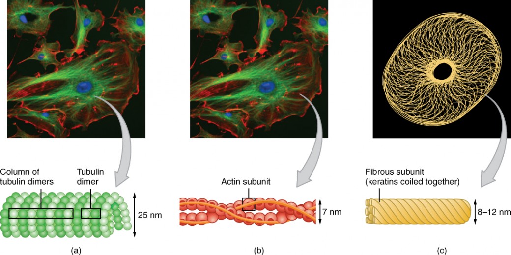
Types of cytoskeletal filaments
Microfilaments (actin filaments) are the thinnest of the three, at about 6–8 nm in diameter. They're composed of actin monomers that polymerize into flexible, two-stranded helical filaments.
- Enable cell movement, including muscle contraction
- Support the plasma membrane and form cellular protrusions like microvilli (which increase surface area for absorption in the intestines) and lamellipodia (sheet-like extensions cells use to crawl)
- Form stress fibers, bundles of actin that provide tension and help anchor cells to surfaces
Intermediate filaments are mid-sized, at 8–12 nm in diameter. Unlike the other two types, they're made from a diverse family of proteins, including keratins (in epithelial cells), lamins (lining the nucleus), and vimentins (in connective tissue cells).
- Provide mechanical strength and resistance to shear stress, helping cells withstand pulling and stretching forces
- Anchor organelles in place within the cytoplasm
- Play key roles in cell-cell and cell-matrix adhesion through structures called desmosomes and hemidesmosomes
- Unlike microfilaments and microtubules, intermediate filaments are not polar and do not serve as tracks for motor proteins
Microtubules are the largest, at 25 nm in diameter. They're hollow, cylindrical structures built from repeating dimers of α-tubulin and β-tubulin.
- Organize the cytoplasm and provide tracks for intracellular transport. Motor proteins called dynein (moves toward the minus end) and kinesin (moves toward the plus end) walk along microtubules carrying cargo like vesicles and organelles.
- Form the mitotic spindle during cell division to pull chromosomes apart
- Provide the structural core of cilia and flagella
- Originate from the centrosome, which acts as the main microtubule-organizing center (MTOC) in animal cells
Cellular Motility and Cytoskeletal Differences
Cilia and flagella in motility
Cilia and flagella are specialized structures that protrude from the cell surface and enable movement. Despite their different sizes and beating patterns, they share the same core architecture.
Structure:
- The internal framework is called the axoneme, arranged in a 9+2 pattern: nine outer doublet microtubules surrounding two central singlet microtubules
- Dynein arms connect the outer doublets to each other and generate the force for bending by using ATP as an energy source
- A basal body at the base anchors the structure to the cell and organizes microtubule assembly. Basal bodies are structurally identical to centrioles.
Function:
- Cilia are shorter and more numerous. They beat in coordinated waves to move fluids or particles across a surface. For example, cilia lining the respiratory tract sweep mucus and trapped debris upward, and cilia in the fallopian tubes help move the egg toward the uterus.
- Flagella are longer and typically fewer in number. They propel entire cells through liquid with a whip-like motion. The classic example is the sperm cell.

Cytoskeleton in prokaryotes vs eukaryotes
Prokaryotic and eukaryotic cells both have internal structural proteins, but the complexity differs significantly.
Prokaryotic cells lack a true cytoskeleton but possess structural homologs of eukaryotic cytoskeletal proteins:
- MreB (an actin homolog) forms a helical filament beneath the cell membrane, helping maintain cell shape and guiding cell wall synthesis
- FtsZ (a tubulin homolog) assembles into a ring called the Z-ring at the cell's midpoint during binary fission, directing where the cell will divide
Eukaryotic cells have the full, complex cytoskeleton with all three filament types. There are notable differences between animal and plant cells:
- Animal cells lack a cell wall, so they rely heavily on the cytoskeleton for structural support and shape. Intermediate filaments are especially important here, providing tensile strength in cells that experience mechanical stress, like skin cells and muscle cells.
- Plant cells have a rigid cell wall that provides most of their structural support and determines their shape. Microtubules are particularly important in plant cells because they guide the deposition of cellulose during cell wall construction. Intermediate filaments are less abundant and less diverse in plant cells compared to animal cells.
Cytoskeleton dynamics and cell polarity
The cytoskeleton is not a fixed structure. It undergoes constant remodeling, with filaments assembling and disassembling in response to the cell's needs. This dynamic behavior is what allows cells to change shape, migrate, and divide.
Cell polarity, the asymmetric organization of a cell (having a distinct "top" and "bottom," for instance), is established and maintained through the arrangement of cytoskeletal elements. Epithelial cells lining your gut, for example, have microvilli on their apical (top) surface but not on their basal (bottom) surface, and the cytoskeleton enforces that difference.
Focal adhesions are another important feature: these are protein complexes where the cytoskeleton (specifically actin filaments) connects to the extracellular matrix outside the cell. They allow cells to grip their surroundings, sense mechanical forces, and transmit signals between the cell's interior and exterior.