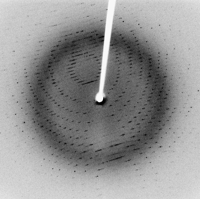
X-ray, electron, and neutron diffraction techniques offer unique insights into crystal structures. Each method has distinct advantages: X-rays excel at heavy elements, electrons provide high resolution for nanostructures, and neutrons are great for light elements and magnetic materials.
Choosing the right technique depends on your sample and research goals. Combining methods can give a more complete picture, from atomic positions to bulk properties. Understanding the strengths of each approach helps scientists pick the best tool for their crystallography needs.
Diffraction Techniques: X-ray vs Electron vs Neutron
Fundamental Principles and Instrumentation
- X-ray diffraction employs electromagnetic radiation with wavelengths comparable to interatomic distances
- Generated by X-ray tubes or synchrotron sources
- Wavelength range typically 0.1 to 100 Å
- Electron diffraction uses high-energy electrons accelerated in an electron microscope
- Wavelengths much shorter than X-rays (0.01 to 0.1 Å)
- Allows study of very small crystalline samples (nanocrystals)
- Neutron diffraction utilizes thermal or cold neutrons
- Produced by nuclear reactors or spallation sources
- Wavelengths similar to X-rays (0.5 to 20 Å)
- X-ray diffraction primarily interacts with electron clouds in atoms
- Neutron diffraction interacts with atomic nuclei
- Provides complementary information on crystal structures
- Electron diffraction demonstrates stronger interaction with matter compared to X-rays and neutrons
- Suitable for studying thin films, nanocrystals, and surface structures
Instrumentation and Facilities
- X-ray diffraction instrumentation includes:
- X-ray sources (sealed tubes, rotating anodes)
- Monochromators
- Goniometers
- Detectors (scintillation counters, area detectors)
- Electron diffraction requires electron microscopes with specialized diffraction attachments
- Transmission electron microscopes (TEM)
- Scanning electron microscopes (SEM) with electron backscatter diffraction (EBSD) capability
- Neutron diffraction facilities are large-scale installations
- Complex shielding requirements due to radiation safety concerns
- Examples include ISIS (UK), ILL (France), and ORNL (USA)
Applications and Scattering Properties
- X-ray diffraction excels in determining atomic positions of heavy elements
- Widely used for overall crystal structure determination
- Applications in pharmaceuticals, materials science, and mineralogy
- Electron diffraction provides high spatial resolution
- Particularly useful for studying nanomaterials and thin films
- Applications in semiconductor industry and nanotechnology
- Neutron diffraction superior for:
- Locating light elements (hydrogen)
- Distinguishing isotopes
- Studying magnetic structures
- Applications in battery research, hydrogen storage materials, and magnetic materials
Strengths and Limitations of Diffraction Techniques

Advantages and Disadvantages of X-ray Diffraction
- Excels in determining atomic positions of heavy elements
- Widely accessible and can be performed in laboratory settings
- Limitations in distinguishing light elements and neighboring elements in the periodic table
- Challenges with highly absorbing or fluorescent materials
- Provides good resolution for overall crystal structures
- Limited penetration depth in some materials
Electron Diffraction Characteristics
- Provides high spatial resolution down to atomic scale
- Useful for studying nanomaterials and thin films
- Suffers from multiple scattering effects
- Complicates quantitative analysis
- Limited penetration depth
- Primarily surface-sensitive technique
- Can provide both structural and chemical information
- When combined with spectroscopic techniques (EELS, EDS)
- Requires very small sample volumes
- Advantageous for limited or precious samples
Neutron Diffraction Features
- Superior for locating light elements (hydrogen, lithium)
- Capable of distinguishing isotopes
- Useful in studying isotope effects in materials
- Excellent for studying magnetic structures
- Spin-dependent scattering
- Requires large sample volumes compared to X-ray and electron diffraction
- Lower resolution compared to X-ray and electron diffraction
- Non-destructive and can penetrate deeply into materials
- Suitable for studying bulk properties
- Enables in-situ experiments (high pressure, high temperature)
Choosing the Right Diffraction Technique
Material Composition Considerations
- Consider elemental composition of the material
- X-rays more suitable for heavy elements (iron, gold)
- Neutrons better for light elements (hydrogen, lithium) and isotope differentiation
- Evaluate sample size and availability
- Electron diffraction requires very small samples (nanograms)
- X-ray diffraction works with moderate amounts (milligrams)
- Neutron diffraction typically needs larger volumes (grams)
- Assess need for spatial resolution
- Electron diffraction offers highest resolution for nanostructures (angstrom scale)
- X-ray diffraction provides good resolution for bulk crystals (nanometer scale)
- Neutron diffraction has lower spatial resolution but probes bulk properties
Specific Structural Information Needs
- Determine if magnetic structure information required
- Neutron diffraction primary choice for magnetic materials (ferromagnets, antiferromagnets)
- Consider time resolution needed for dynamic studies
- Synchrotron X-ray sources provide high temporal resolution (picoseconds)
- Pulsed neutron sources offer good time resolution (microseconds)
- Evaluate need for bulk vs. surface structural information
- X-rays and neutrons probe bulk properties
- Electrons more surface-sensitive (nanometers depth)
- Assess requirement for in-situ or operando studies
- Neutron diffraction advantageous due to penetrating power
- Allows studies under extreme conditions (high pressure, high temperature)
Analyzing Complementary Diffraction Data
Combining X-ray and Neutron Diffraction
- Integrate X-ray and neutron diffraction data to accurately determine both heavy and light atom positions
- X-rays locate heavy atoms (metal centers)
- Neutrons pinpoint light atoms (hydrogen bonding networks)
- Combine X-ray and neutron powder diffraction results
- Resolve structural ambiguities
- Improve overall structural model accuracy
- Use X-ray and neutron pair distribution function (PDF) analysis together
- Probe short-range and long-range order
- Applicable to both crystalline and amorphous materials
Integrating Electron Diffraction with Other Techniques
- Utilize electron diffraction to supplement X-ray or neutron data
- Provide high-resolution information on local structure and defects
- Correlate single-crystal X-ray diffraction with electron diffraction tomography
- Bridge gap between atomic and nanoscale structural features
- Combine electron diffraction with spectroscopic methods
- Electron energy loss spectroscopy (EELS) for elemental mapping
- Energy-dispersive X-ray spectroscopy (EDS) for chemical composition
Multi-technique Approach for Comprehensive Analysis
- Combine neutron diffraction data with X-ray or electron diffraction
- Simultaneously analyze nuclear and magnetic structures
- Integrate information from multiple diffraction techniques
- Validate and refine structural models
- Ensure consistency across different length scales
- Provide comprehensive view from atomic to bulk properties
- Use complementary techniques to overcome individual limitations
- X-ray diffraction for overall structure
- Neutron diffraction for light elements and magnetism
- Electron diffraction for local structure and nanofeatures