Laue Method Principles
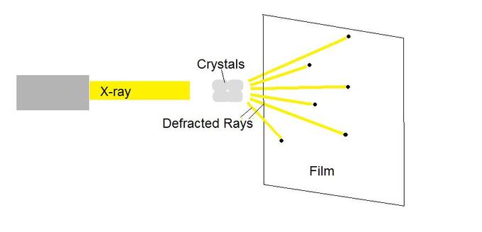
Experimental Setup and Diffraction Process
The Laue method is the oldest X-ray diffraction technique, and it works on a beautifully simple idea: instead of tuning a single wavelength to find diffraction conditions, you flood a stationary crystal with all wavelengths at once and let the crystal pick out which ones diffract. This polychromatic (or "white beam") approach means every set of lattice planes can find some wavelength in the beam that satisfies Bragg's law, producing a rich pattern of spots in a single exposure.
The experimental setup has four main components:
- X-ray source producing a continuous spectrum of wavelengths (polychromatic beam)
- Collimator to shape the beam into a narrow, well-defined pencil
- Stationary single crystal as the sample
- Flat film or area detector positioned perpendicular to the incident beam
Each spot in the resulting Laue pattern corresponds to a different set of lattice planes (with Miller indices ), each diffracting at whatever wavelength from the beam happens to satisfy the Bragg condition for that plane's spacing and orientation.
Two main geometries are used:
- Transmission (forward-scattering): The detector sits behind the crystal. This works for thin samples or materials with low X-ray absorption. A typical use is studying thin film crystal orientations.
- Back-reflection (back-scattering): The detector sits between the X-ray source and the crystal, with a hole for the beam to pass through. This is better for thick or highly absorbing samples, such as analyzing surface structures of bulk metals.
Your choice between these depends on sample thickness, absorption coefficient, and what information you need.
Mathematical Foundations and Geometrical Considerations
Bragg's law governs the diffraction process:
where is the integer order of reflection, is the X-ray wavelength, is the interplanar spacing for the planes, and is the Bragg angle. In the Laue method, is fixed by the crystal orientation, and varies continuously, so diffraction occurs at whatever wavelength satisfies this equation for each plane.
The Ewald sphere construction helps visualize this in reciprocal space. The Ewald sphere has radius . Because the Laue method uses a range of wavelengths ( to ), you get a range of Ewald sphere radii, forming a thick shell. Any reciprocal lattice point falling between the spheres of radius and will produce a diffraction spot. That's why Laue patterns contain so many reflections in a single shot.
The Laue equations describe the diffraction geometry in terms of the unit cell axes directly:
Here , , are unit cell parameters; are angles between the diffracted beam and the unit cell axes; are angles between the incident beam and the unit cell axes; and are Miller indices. All three equations must be satisfied simultaneously for diffraction to occur, which is equivalent to satisfying Bragg's law.
Gnomonic projection is a geometrical tool for analyzing Laue patterns. It projects diffraction spots onto a plane tangent to a reference sphere, which straightens out the curved zone lines (see below) into straight lines. This makes it much easier to identify zone axes and measure interplanar angles.
Interpreting Laue Patterns
Symmetry Analysis and Crystal Orientation Determination
A central feature of Laue patterns is that the arrangement of spots reflects the point group symmetry of the crystal along the beam direction. If you shoot the beam along a 4-fold axis of a cubic crystal, the pattern will show 4-fold symmetry. This direct connection between pattern symmetry and crystal symmetry is what makes the Laue method so powerful for orientation determination.
Key aspects of pattern interpretation:
- The distribution and relative intensity of spots encode information about the crystal's lattice parameters and atomic arrangement.
- Systematic absences reveal space group symmetry. For example, in a body-centered cubic structure, reflections where is odd are absent. Spotting these absences helps you narrow down the space group.
- Spots in a Laue pattern fall along curves called zone lines, each corresponding to a crystallographic zone axis. Identifying these zones is the first step in determining orientation.
Indexing a Laue pattern means assigning Miller indices to each spot. This can be done:
- Manually, using Greninger charts or stereographic projections to map spot positions to crystal directions
- Automatically, using software such as LaueGen or CLIP, which is faster and less error-prone
Stereographic projections compress the 3D crystal orientation into a 2D map, making it straightforward to identify zone axes and calculate angles between crystal planes. The gnomonic projection serves a similar purpose but is especially convenient for Laue geometry because zone lines project as straight lines.
Advanced Computational Techniques
Modern Laue analysis relies heavily on computation:
- Automated indexing algorithms (e.g., Hough transform-based methods) detect spots and assign indices rapidly, enabling real-time orientation determination.
- Pattern matching compares experimental patterns against simulated ones from known structures to confirm or refine orientation.
- Machine learning approaches, such as convolutional neural networks, are increasingly used for automated pattern classification, especially when dealing with large datasets.
- Quantitative intensity analysis can estimate crystal mosaicity (the spread of orientations within a nominally single crystal) and lattice distortions. Rocking curve analysis, where the crystal is tilted slightly while monitoring a reflection, provides a direct measure of crystal quality.
- Multi-technique integration combines Laue diffraction with X-ray fluorescence (for elemental analysis) or X-ray topography (for imaging defects) to build a more complete picture of the crystal.
Laue Method Applications

Crystal Perfection and Defect Analysis
The shape and sharpness of Laue spots are direct indicators of crystal quality:
| Spot Feature | What It Indicates |
|---|---|
| Sharp, round spots | High crystal perfection |
| Diffuse or broadened spots | Imperfections, mosaic spread, or disorder |
| Asterism (elongated/streaked spots) | Crystal deformation or internal strain (e.g., plastic deformation in metals under stress) |
| Split spots | Twinning or low-angle grain boundaries (e.g., twin structures in shape memory alloys) |
| Extra spots or diffuse scattering between main reflections | Short-range order, disorder, or secondary phases (e.g., order-disorder transitions in alloys) |
| Continuous streaks | Stacking faults or other planar defects (e.g., stacking faults in semiconductor materials) |
This makes the Laue method a quick diagnostic tool: a single exposure can tell you whether a crystal is high-quality, strained, twinned, or contains secondary phases.
Dynamic Studies and Advanced Applications
Because Laue patterns can be collected in a single, short exposure (no crystal rotation needed), the method is well suited for time-resolved and in situ studies:
- Time-resolved Laue diffraction captures structural changes as they happen, on timescales down to picoseconds at synchrotron sources. This is used to investigate phase transitions in ferroelectric materials or track photo-induced structural dynamics.
- In situ studies under extreme conditions include high-pressure experiments in diamond anvil cells and high-temperature studies of refractory materials, where the crystal structure is monitored while conditions change.
- Micro-Laue diffraction uses a focused beam (micron-scale) to map strain fields and orientation variations across polycrystalline or heterogeneous samples. For example, scanning a focused white beam across a polycrystalline metal sample produces an orientation and strain map with spatial resolution.
- Protein crystallography uses Laue diffraction for rapid screening of crystal quality and orientation before committing to full monochromatic data collection. It's also used for time-resolved studies of enzyme reactions.
- Multi-modal analysis combines Laue diffraction with spectroscopic techniques (e.g., simultaneous X-ray absorption spectroscopy) to probe both local electronic structure and long-range crystalline order in the same experiment.
Laue Method: Advantages vs. Limitations
Advantages
- Speed: Crystal orientation can be determined in a single exposure with no need for precise sample alignment or monochromatic radiation. This makes it valuable for quick quality checks in industrial and research settings.
- Comprehensive reciprocal space coverage: A single shot samples many reflections simultaneously, giving an efficient overview of crystal quality. Protein crystallographers use this for rapid screening before detailed data collection.
- Large crystal analysis: Particularly useful for studying large single crystals in mineralogy and materials science (e.g., characterizing gemstones and mineral specimens).
- Non-destructive and in situ capable: The crystal isn't consumed or damaged (barring radiation effects), and measurements can be performed while the sample is heated, cooled, or pressurized.
- Time-resolved capability: The single-exposure nature makes it ideal for studying fast structural dynamics in photo-activated or stimuli-responsive materials.
Limitations
- No precise lattice parameters from a single pattern. Because each spot diffracts at a different (unknown) wavelength, you can't directly extract unit cell dimensions the way you can with monochromatic methods. You get orientation and symmetry, but not accurate -spacings without additional calibration.
- Overlapping reflections. Different orders of the same reflection (e.g., and ) diffract at different wavelengths but land on the same spot, complicating intensity analysis. This is especially problematic for low-symmetry crystals like monoclinic or triclinic systems.
- Not suited for polycrystalline samples. The method is designed for single crystals. It can't replace powder diffraction for polycrystalline materials, and quantitative structure factor determination is difficult because intensities are integrated over multiple wavelengths.
- Radiation damage. The intense white beam delivers more total radiation than a monochromatic beam, which can degrade sensitive samples. Protein crystals, for instance, may suffer significant damage during extended exposures.
- Complex data analysis. Interpreting Laue patterns, especially for quantitative work, requires specialized software and expertise that go beyond standard monochromatic diffraction analysis.
- Limited quantitative phase analysis. In multi-phase systems, distinguishing between phases with similar structures is difficult because of the wavelength ambiguity inherent in the method.