The Human Visual System
Vision starts with light entering the eye and ends with your brain constructing a meaningful image from electrical signals. Understanding how this chain works, from eye structures to brain processing, is central to the sensation and perception unit.
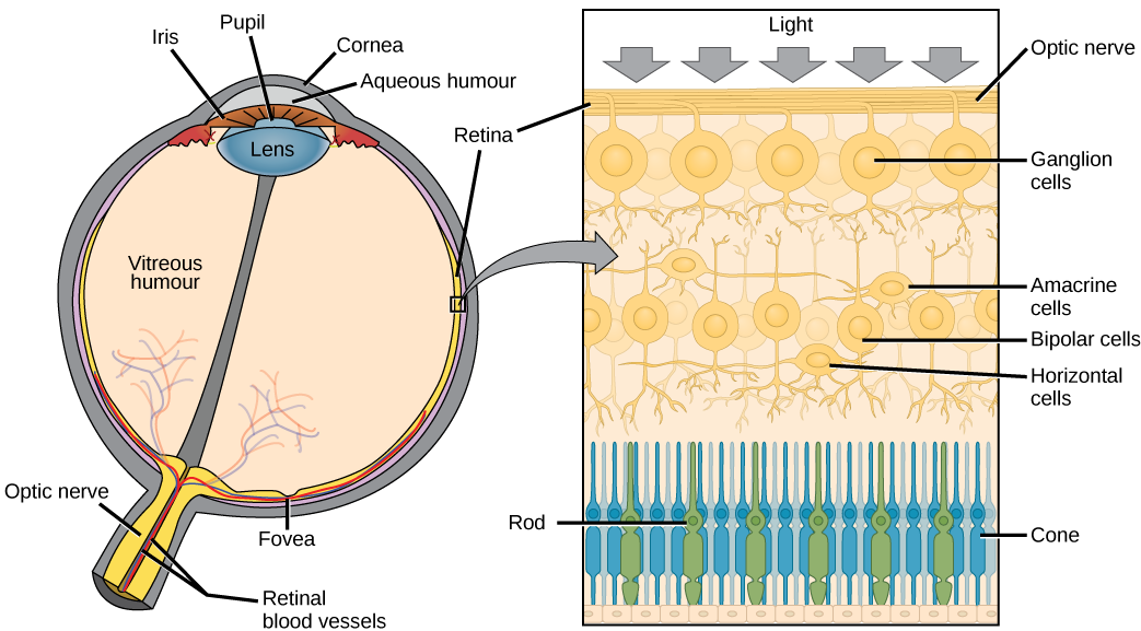
Components of the Human Visual System
Light passes through several structures in the eye before it ever becomes a neural signal. Each structure plays a specific role:
- Cornea: the transparent outer layer that bends (refracts) light as it enters the eye. Most of the eye's focusing power comes from the cornea.
- Pupil: the opening in the center of the iris that lets light pass through.
- Iris: the colored ring of muscle surrounding the pupil. It controls how much light enters by making the pupil larger (dilating) or smaller (constricting).
- Lens: sits behind the pupil and fine-tunes focus by changing shape, a process called accommodation. It flattens for distant objects and thickens for close ones.
- Retina: the light-sensitive layer at the back of the eye, containing photoreceptor cells (rods and cones) that convert light into neural signals. This is where transduction happens.
Once the retina converts light into neural impulses, those signals travel along the optic nerve to the brain. The signals pass through the lateral geniculate nucleus (LGN) of the thalamus and then reach the visual cortex in the occipital lobe.
- Primary visual cortex (V1): the first cortical area to receive visual input from the LGN. Neurons here, called feature detectors, respond to specific elements like edges, lines, and angles.
- Higher visual areas (V2, V3, V4, V5): process increasingly complex features such as form, color, motion, and depth. V4, for example, is heavily involved in color processing, while V5 handles motion.
Rod vs. Cone Cells
The retina has two types of photoreceptors, and they do very different jobs.
Rod cells handle low-light vision:
- Extremely sensitive to light, making them essential for seeing at night or in dim rooms (scotopic vision)
- Contain a photopigment called rhodopsin that responds to even small amounts of light
- Concentrated in the peripheral (outer) regions of the retina, which is why you can sometimes see a faint star better by looking slightly to the side of it
- Do not detect color; they only register light intensity (shades of gray)
- There are roughly 120 million rods in each eye
Cone cells handle color and detail:
- Less sensitive to light, so they work best in well-lit conditions (photopic vision)
- Come in three types, each tuned to a different range of wavelengths:
- S-cones: short wavelengths (blue)
- M-cones: medium wavelengths (green)
- L-cones: long wavelengths (red)
- Your brain perceives color by comparing the relative activation across these three cone types. Seeing yellow, for instance, involves strong activation of both L-cones and M-cones.
- Concentrated in the fovea, the small central pit of the retina responsible for sharp, detailed vision. This is why you look directly at something when you want to see it clearly.
- There are roughly 6 million cones in each eye
Depth Perception
Depth perception is your ability to see the world in three dimensions even though the image on each retina is flat (two-dimensional). Your brain uses two categories of cues to pull this off.
Monocular vs. Binocular Depth Cues
Monocular cues work with just one eye. These are the same cues artists use to create the illusion of depth on a flat canvas:
- Relative size: objects that take up less space on your retina are perceived as farther away. A person across a football field looks tiny compared to someone standing next to you.
- Interposition (occlusion): when one object partially blocks another, the blocked object is seen as farther away. A fence post in front of a house tells you the post is closer.
- Linear perspective: parallel lines appear to converge as they stretch into the distance. Think of railroad tracks seeming to meet at a point on the horizon.
- Texture gradient: surface detail becomes finer and more compressed with distance. Blades of grass look distinct near your feet but blend into a smooth green carpet farther out.
- Aerial perspective: distant objects look hazier and more washed out because of particles in the atmosphere. Far-off mountains often appear bluish or faded.
- Motion parallax: when you're moving, nearby objects seem to zip past quickly while distant objects barely move. Watch the difference between roadside signs and distant hills from a car window.
Binocular cues require both eyes working together:
- Binocular disparity (retinal disparity): because your eyes are a few centimeters apart, each one receives a slightly different image. Your brain fuses these two images into a single three-dimensional percept, a process called stereopsis. 3D movies exploit this by sending a different image to each eye.
- Convergence: your eyes angle inward when focusing on something close. The brain uses the degree of this inward rotation as a distance cue. You can feel convergence if you try to focus on your fingertip as you slowly bring it toward your nose.

Visual Processing and Perception
Perceptual Organization
Raw visual input is a flood of light, edges, and colors. Your brain has to organize all of that into objects and scenes you can actually make sense of. Several principles explain how it does this.
Gestalt principles describe the rules your brain follows when grouping visual elements. The core idea is that the whole percept is different from the sum of its parts. Key principles include:
- Proximity: elements close together are grouped as a unit
- Similarity: elements that look alike (same color, shape, or size) are grouped together
- Closure: your brain fills in gaps to perceive a complete shape, even when parts are missing
- Continuity: you tend to perceive smooth, continuous lines rather than abrupt changes in direction
Figure-ground separation is how you distinguish an object (the figure) from its background (the ground). This usually happens automatically, but ambiguous images like Rubin's vase show that your brain can flip between two interpretations.
Perceptual constancies keep your experience of objects stable even when the sensory input changes:
- Size constancy: a friend walking away from you doesn't seem to shrink, even though their image on your retina gets smaller
- Shape constancy: a door looks rectangular whether it's closed (rectangle on your retina) or half-open (trapezoid on your retina)
- Color constancy: a red apple still looks red under fluorescent lighting, sunlight, or candlelight, even though the wavelengths reaching your eye differ in each case
Visual Field and Processing
The visual field is the entire area you can see when looking straight ahead, including your peripheral vision. Information from the left visual field goes to the right hemisphere, and information from the right visual field goes to the left hemisphere. This crossover happens at the optic chiasm, where fibers from the nasal (inner) half of each retina cross to the opposite side of the brain.
The brain processes different aspects of a visual scene in parallel. Color, motion, form, and depth are handled by specialized areas and then integrated so you experience one unified image rather than separate fragments. This binding of features into a coherent whole is still an active area of research in psychology and neuroscience.