Viral capsids are the protein shells that protect a virus's genetic material. They come in different shapes and sizes, with icosahedral (spherical) and helical (rod-shaped) being the main types. These structures play a crucial role in how viruses infect cells and survive outside their hosts.

Capsids are made up of smaller units called capsomeres, which fit together like puzzle pieces. The way these pieces arrange determines the capsid's shape and affects how the virus interacts with host cells, evades the immune system, and withstands environmental stresses.

Viral Capsid Structure

Protein Shell Composition and Function

Top images from around the web for Protein Shell Composition and Function
  • Virus capsids form protein shells enclosing and protecting viral genomes
  • Capsids comprise multiple copies of protein subunits called capsomeres
  • Capsomere arrangement determines overall capsid geometric shape
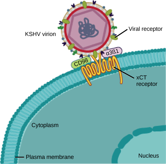
  • Capsid proteins often contain specific binding sites for host cell receptors
    • These binding sites play a crucial role in viral attachment and entry
  • Some viruses have additional structural features
    • Lipid envelope surrounding the capsid
    • Protein spikes protruding from capsid surface

Capsid Size and Diversity

  • Viral capsid size ranges from ~20 to 400 nanometers in diameter
  • Larger viruses generally have more complex capsid structures
  • Two main categories of capsids based on symmetry
    • Icosahedral (spherical)
    • Helical (rod-shaped)
  • Capsid structure varies among virus types
    • Influences viral properties and behavior

Icosahedral vs Helical Symmetry

Structural Characteristics

  • Icosahedral capsids
    • Spherical or near-spherical shape
    • 20 triangular faces and 12 vertices
    • Composed of pentamers and hexamers of capsid proteins
    • More rigid structure
  • Helical capsids
    • Rod-like or filamentous structure
    • Formed by a single type of capsomere arranged in a spiral
    • More flexible structure, allowing variation in length

Assembly and Genome Packing

    • Allows efficient packing of subunits to enclose spherical space
    • Assembly often occurs from preformed capsomere clusters
    • More suitable for enclosing linear genomes
    • Forms by sequential addition of subunits to one end
  • Icosahedral capsids common in DNA viruses and some RNA viruses
  • Helical capsids predominantly found in RNA viruses (particularly single-stranded)

Capsid Structure and Viral Function

Environmental Stability and Protection

  • Capsid structure influences virus's ability to withstand environmental stresses
    • Temperature changes
    • pH variations
    • Enzymatic degradation
  • Capsid stability crucial for
    • Protecting viral genome during transmission between hosts
    • Maintaining infectivity outside host cell

Host Cell Interaction and Entry

  • Capsid protein arrangement determines positioning of receptor binding sites
    • Affects virus's ability to recognize and attach to host cells
  • Capsid structure influences mechanism of viral entry (endocytosis, membrane fusion)
  • Some capsids allow conformational changes upon host cell interaction
    • Facilitates release of viral genome into host cell

Immune Evasion and Replication

  • Certain capsid structures can evade or modulate host immune responses
    • Contributes to virus's overall infectivity and persistence
  • Capsid structural integrity essential for proper assembly and disassembly
    • Crucial during viral replication cycle

Capsomere Types and Assembly

Basic Capsomere Structures

  • Capsomeres serve as basic building blocks of viral capsids
  • Typically composed of multiple copies of one or more capsid proteins
  • Common capsomere types
    • Pentamers (five protein subunits)
    • Hexamers (six protein subunits)
    • Monomers (single protein subunits in helical capsids)
  • Specialized capsomeres in some viruses
    • Trimers (three protein subunits)
    • Dimers (two protein subunits)

Capsomere Roles and Interactions

  • Pentamers often found at vertices of icosahedral capsids
    • Crucial for initiating capsid curvature
  • Hexamers form faces of icosahedral capsids
    • Contribute to overall capsid structure
  • In helical capsids, single protein subunits polymerize to form helical structure
  • Capsomeres interact through non-covalent bonds
    • Allows for reversible assembly and disassembly of capsid
  • Arrangement and interactions between capsomere types determine
    • Final capsid geometry
    • Overall capsid stability

Key Terms to Review (18)

Adenovirus: Adenoviruses are a group of medium-sized, non-enveloped viruses that are known to cause a variety of illnesses in humans and animals, particularly respiratory infections, conjunctivitis, and gastroenteritis. They play a significant role in viral taxonomy, structural biology, and mechanisms of infection.
Capsid assembly: Capsid assembly is the process by which viral proteins, specifically capsomers, come together to form the protective protein shell known as the capsid around the viral genome. This assembly is crucial for the virus's stability, infectivity, and ability to evade host immune responses. Understanding capsid assembly helps to explain how viruses achieve structural symmetry and stability while also linking to the functions performed by various viral proteins during this critical stage.
Capsomers: Capsomers are the individual protein subunits that make up a viral capsid, the protective outer shell of a virus. They play a crucial role in the overall structure and stability of the capsid, which is essential for the virus's ability to infect host cells. By organizing into specific geometric arrangements, capsomers contribute to the symmetry of the viral capsid, impacting how the virus interacts with its environment and host organisms.
Electron microscopy: Electron microscopy is a powerful imaging technique that uses a beam of electrons to visualize specimens at a very high resolution, allowing scientists to see structures at the nanometer scale. This technique is essential for studying viruses, as it provides detailed images of viral capsids, helps to identify different viral structures, and aids in understanding complex processes like virion assembly and maturation.
Enveloped viruses: Enveloped viruses are a type of virus that have an outer lipid membrane, known as an envelope, surrounding their capsid. This envelope is derived from the host cell membrane during the budding process of viral replication, which provides the virus with a unique means of entry into host cells and enhances its ability to evade the immune system. Understanding enveloped viruses helps connect important concepts such as viral capsid structures, symmetry, and their roles in various virus families.
Facilitation of viral entry: Facilitation of viral entry refers to the mechanisms by which viruses attach to and penetrate host cells, allowing them to deliver their genetic material and initiate infection. This process involves specific interactions between viral capsid structures and host cell receptors, with the geometry and symmetry of the viral capsid playing critical roles in determining how effectively a virus can bind to and enter a host cell.
Helical capsid: A helical capsid is a type of viral protein shell that has a cylindrical or spiral shape, where the protein subunits are arranged in a helical pattern around the viral nucleic acid. This structure not only provides protection to the viral genome but also plays a crucial role in the virus's ability to infect host cells. Helical capsids are typically found in viruses with single-stranded RNA genomes, and their symmetry contributes to the overall stability and functionality of the virus.
Helical symmetry: Helical symmetry refers to a structural arrangement found in certain viruses where the capsid proteins are arranged in a spiral or helical pattern around the viral nucleic acid. This organization allows for the efficient packaging of the viral genome and provides a sturdy protective structure. Helical symmetry is a key feature that influences how viruses are classified and how they interact with host cells.
Icosahedral capsid: An icosahedral capsid is a virus structure characterized by a geometric shape that has 20 equilateral triangular faces, making it highly symmetrical and efficient for enclosing viral genetic material. This structure allows for a compact and stable assembly, which is crucial for the protection and delivery of the viral genome during infection. The symmetry of an icosahedral capsid contributes to the stability and resilience of the virus against environmental stresses.
Icosahedral symmetry: Icosahedral symmetry is a form of molecular symmetry exhibited by certain viruses, characterized by a geometric structure that resembles an icosahedron. This symmetry allows viruses to efficiently enclose their genetic material in a protective shell, optimizing stability and structural integrity. This design is crucial in determining how viruses interact with host cells, influencing both their classification and characteristics.
Naked Viruses: Naked viruses are viral particles that lack an outer lipid envelope, consisting only of a nucleic acid genome encased in a protein shell called a capsid. This absence of an envelope gives naked viruses unique structural properties and affects their mode of transmission, resistance to environmental factors, and interactions with host cells.
Nucleocapsid: A nucleocapsid is the structural complex formed by the combination of a viral genome and its protective protein coat, known as the capsid. This assembly plays a crucial role in protecting the viral genetic material and facilitating its delivery into host cells, linking it to important features such as viral structure, genome organization, and replication processes.
Protection of viral genome: Protection of viral genome refers to the mechanisms by which viruses safeguard their genetic material from degradation and environmental threats. This is crucial for the survival and replication of viruses, ensuring that their genetic information remains intact during transmission between hosts and within host cells. The structural features of a virus, such as the viral capsid, play a significant role in this protection by encapsulating the genome and providing a stable environment for its integrity.
Structure-Function Relationship: The structure-function relationship refers to the concept that the specific arrangement and organization of components within a biological entity, such as a virus, determines its functions and interactions. This idea is particularly relevant in virology, where the architecture of viral capsids influences how viruses infect host cells, protect their genetic material, and determine their overall stability and infectivity.
Tobacco mosaic virus: Tobacco mosaic virus (TMV) is a rod-shaped plant virus that infects a wide range of plant species, particularly tobacco and other members of the Solanaceae family. It was the first virus to be discovered and characterized, making it a foundational element in the history of virology and significantly contributing to our understanding of viral structure and behavior.
Viral maturation: Viral maturation is the final stage in the viral life cycle, where newly formed viral particles undergo structural changes to become infectious. This process involves the assembly and modification of the viral capsid and other components, ensuring that the virus is stable and capable of successful infection upon release from the host cell. The arrangement of proteins in the capsid plays a crucial role in defining the virus's symmetry and stability, which are essential for its ability to infect new host cells.
Viral stability: Viral stability refers to the ability of a virus to maintain its structure and function over time and under varying environmental conditions. This concept is crucial because the stability of a virus influences its infectivity, transmission, and overall survival outside of a host organism. Factors such as temperature, pH, and the presence of solvents can significantly impact viral stability, making it an important consideration in virology.
X-ray Crystallography: X-ray crystallography is a powerful analytical technique used to determine the atomic and molecular structure of a crystal by diffracting X-rays through it. This method reveals how the atoms are arranged in a crystal lattice, providing detailed insights into the structure of biological macromolecules, such as viral capsids, and their symmetry, which is crucial for understanding how viruses assemble and function.
© 2024 Fiveable Inc. All rights reserved.
AP® and SAT® are trademarks registered by the College Board, which is not affiliated with, and does not endorse this website.