🫁Honors Anatomy and Physiology Unit 11 Review
11.1 Lymphatic System Structure and Function
11.1 Lymphatic System Structure and Function
Unit & Topic Study Guides
Intro to Anatomy & Physiology
Chemical Basis of Life
Cellular Structure and Function
Integumentary System
Skeletal System
Muscular System
Nervous System
Cardiovascular System
Lymphatic & Immune Systems
Respiratory System
Digestive System
Urinary System
Reproductive System
Development and Inheritance
Homeostasis and Regulation
Advanced Anatomy & Physiology Topics
The lymphatic system is your body's unsung hero, working tirelessly to keep you healthy. It's a network of vessels and organs that filters out bad stuff, fights infections, and helps absorb fats from your food.
Lymph nodes, spleen, and thymus are key players in this system. They're like training camps for immune cells, getting them ready to fight off invaders. The lymphatic system also helps maintain fluid balance, preventing swelling in your body.
Lymphatic System Anatomy and Function

Lymphatic Vessels and Lymph Composition
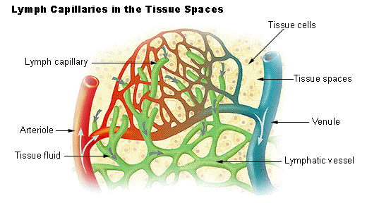
- The lymphatic system is a network of vessels, tissues, and organs that helps maintain fluid balance, fight infection, and absorb fats from the digestive tract
- Lymphatic vessels are thin-walled, valved structures that carry lymph, a clear fluid containing white blood cells, proteins, and lipids
- Lymphatic capillaries are highly permeable, allowing for the uptake of interstitial fluid and the formation of lymph
- Lymphatic vessels progressively increase in size, from lymphatic capillaries to collecting vessels, and ultimately drain into the subclavian veins via the thoracic duct and right lymphatic duct
Lymphoid Organs and Their Functions
- Lymphoid organs, including lymph nodes, the spleen, the thymus, and mucosa-associated lymphoid tissue (MALT), are sites where immune cells congregate and mount immune responses
- The lymphatic system plays a crucial role in maintaining fluid balance by returning excess interstitial fluid to the bloodstream, preventing edema (swelling)
- Lymphatic vessels in the small intestine, called lacteals, absorb dietary fats and fat-soluble vitamins (Vitamins A, D, E, and K), transporting them as chyle to the bloodstream via the thoracic duct
- The lymphatic system also facilitates the transport of immune cells, such as lymphocytes, and antigen-presenting cells (dendritic cells) throughout the body, enabling effective immune surveillance and response
Lymph Node Filtering and Immunity
Lymph Node Structure and Lymph Filtration
- Lymph nodes are small, bean-shaped structures located along lymphatic vessels that filter lymph and provide a site for immune cell activation and proliferation
- Lymph enters the lymph node through afferent lymphatic vessels, passes through a series of sinuses, and exits via efferent lymphatic vessels
- Macrophages and dendritic cells within the lymph node remove pathogens, debris, and abnormal cells from the lymph, preventing their spread to other parts of the body
- High endothelial venules (HEVs) in lymph nodes allow for the continuous migration of lymphocytes from the bloodstream into the lymph node, ensuring a constant supply of immune cells
Lymphocyte Activation and Differentiation in Lymph Nodes
- Lymph nodes contain specialized compartments, such as the cortex and medulla, which are populated by B cells and T cells, respectively
- B cells in the cortex can encounter antigens and differentiate into plasma cells, which secrete antibodies (immunoglobulins) specific to the encountered antigen
- T cells in the paracortex and medulla can be activated by antigen-presenting cells, such as dendritic cells, and differentiate into effector T cells (helper T cells and cytotoxic T cells)
- Enlarged or tender lymph nodes can indicate an active immune response to infection or inflammation in the area drained by the affected lymph nodes
- Lymph node activation and the subsequent immune response help to contain and eliminate pathogens, preventing systemic spread and disease
Lymphatic vs Blood Vessels

Structural and Functional Differences
- While both lymphatic vessels and blood vessels are involved in circulation, they have distinct functions and structural differences
- Blood vessels, consisting of arteries, capillaries, and veins, transport oxygenated blood and nutrients to tissues and remove deoxygenated blood and waste products
- Blood flows in a closed circuit, propelled by the pumping action of the heart
- Blood capillaries are selectively permeable, allowing for the exchange of gases (oxygen and carbon dioxide), nutrients, and waste products between the blood and tissues
- Lymphatic vessels, in contrast, form an open, one-way system that carries lymph from tissues back to the bloodstream
- Lymphatic vessels have a thinner wall structure compared to blood vessels and lack a central pump. Instead, lymph flow is facilitated by the contraction of skeletal muscles and the presence of one-way valves
- Lymphatic capillaries are more permeable than blood capillaries, allowing for the uptake of larger molecules, such as proteins (albumin), and pathogens that have escaped the bloodstream
Roles in Fluid Balance and Immune Function
- Lymphatic vessels play a crucial role in maintaining fluid balance by returning excess interstitial fluid to the bloodstream, whereas blood vessels are primarily responsible for the transport of gases, nutrients, and waste products
- While blood vessels deliver immune cells and antibodies to sites of infection or inflammation, lymphatic vessels transport antigens and activated immune cells to lymph nodes, facilitating the development of an adaptive immune response
- The lymphatic system complements the functions of the cardiovascular system by managing fluid homeostasis and supporting immune surveillance and response throughout the body
Major Lymphatic Organs and Roles
Lymph Nodes and Spleen
- The major lymphatic organs include the lymph nodes, spleen, thymus, and mucosa-associated lymphoid tissue (MALT), each with specific roles in the immune system
- Lymph nodes are strategically located throughout the body and serve as filters for lymph and sites for immune cell activation and proliferation
- Lymph nodes trap and remove pathogens (bacteria and viruses), debris, and abnormal cells (cancer cells) from the lymph
- They provide a microenvironment for B cell and T cell activation and differentiation, facilitating the development of adaptive immune responses (humoral and cell-mediated immunity)
- The spleen is the largest lymphoid organ and plays a critical role in filtering blood and mounting immune responses against blood-borne pathogens
- The white pulp of the spleen contains B cell follicles and T cell zones, similar to lymph nodes, allowing for the activation and differentiation of lymphocytes
- The red pulp of the spleen removes old or damaged red blood cells (erythrocytes) and platelets (thrombocytes) from circulation, and it can also serve as a reservoir for blood
Thymus and Mucosa-Associated Lymphoid Tissue (MALT)
- The thymus is a primary lymphoid organ located in the upper chest, responsible for the development and maturation of T cells
- Immature T cells, called thymocytes, undergo a selection process in the thymus to ensure they can recognize foreign antigens presented by the body's own cells without reacting to self-antigens (self-tolerance)
- The thymus is most active during childhood and undergoes involution with age, but it continues to play a role in T cell development throughout life
- Mucosa-associated lymphoid tissue (MALT) is a collection of lymphoid tissues found in mucous membranes lining the digestive, respiratory, and urogenital tracts
- MALT includes Peyer's patches in the small intestine, tonsils, and adenoids, among other tissues
- These tissues serve as a first line of defense against pathogens entering the body through mucosal surfaces (oral cavity, respiratory tract, and gastrointestinal tract), inducing both local and systemic immune responses (secretory IgA production)
- The coordinated functions of these lymphatic organs ensure effective immune surveillance, pathogen elimination, and the development of immunological memory, providing long-lasting protection against future encounters with the same pathogens