X-ray diffraction is a powerful technique for determining the atomic structure of crystals. It uses X-rays to scatter off atoms, creating patterns that reveal how they're arranged. This method is crucial in nanobiotechnology for studying biomolecules and nanomaterials.
Understanding X-ray diffraction starts with electromagnetic radiation basics and Bragg's law. These principles explain how X-rays interact with matter and the conditions for diffraction. Various techniques like powder and single-crystal diffraction are used, each with specific applications in nanobiotechnology.
Principles of X-ray diffraction
- X-ray diffraction is a powerful analytical technique used to determine the atomic and molecular structure of crystalline materials, including biological macromolecules and nanomaterials
- The principles of X-ray diffraction are based on the interaction of electromagnetic radiation with matter, specifically the scattering of X-rays by the electron clouds surrounding atoms in a crystal lattice
- Understanding the fundamentals of X-ray diffraction is crucial for its application in nanobiotechnology, as it enables the characterization of biomolecules and nanomaterials at the atomic level
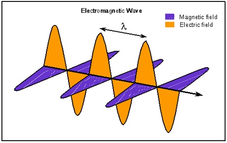
Electromagnetic radiation fundamentals
- Electromagnetic radiation consists of oscillating electric and magnetic fields that propagate through space as waves
- X-rays are a form of electromagnetic radiation with wavelengths ranging from 0.01 to 10 nanometers, positioned between ultraviolet light and gamma rays in the electromagnetic spectrum
- The properties of X-rays, such as their short wavelength and high energy, make them suitable for probing the atomic structure of materials
Wavelength vs photon energy
- The energy of a photon is inversely proportional to its wavelength, as described by the equation , where is the photon energy, is Planck's constant, is the speed of light, and is the wavelength
- X-rays used in diffraction experiments typically have energies ranging from a few keV to tens of keV, corresponding to wavelengths of a few angstroms (0.1 nm)
- The choice of X-ray wavelength depends on the size of the unit cell and the desired resolution of the diffraction data
Interaction of X-rays with matter
- When X-rays interact with matter, they can be absorbed, transmitted, or scattered by the atoms in the material
- Scattering occurs when X-rays interact with the electron clouds surrounding the atoms, causing the electrons to oscillate and emit secondary X-rays in various directions
- The intensity and direction of the scattered X-rays depend on the arrangement of atoms in the crystal lattice and the electron density distribution within the unit cell
Bragg's law
- Bragg's law is a fundamental principle in X-ray diffraction that describes the conditions necessary for constructive interference of scattered X-rays, leading to the formation of diffraction patterns
- The law relates the wavelength of the X-rays, the spacing between lattice planes, and the angle at which constructive interference occurs
- Bragg's law is essential for interpreting X-ray diffraction data and determining the crystal structure of materials
Derivation of Bragg's equation
- Bragg's equation is derived by considering the path difference between X-rays scattered from adjacent lattice planes
- For constructive interference to occur, the path difference must be an integer multiple of the X-ray wavelength
- The equation is expressed as , where is an integer (order of reflection), is the X-ray wavelength, is the spacing between lattice planes, and is the angle of incidence and reflection
Lattice planes and Miller indices
- Lattice planes are imaginary planes that pass through the points of a crystal lattice, describing the periodic arrangement of atoms in the crystal
- Miller indices (hkl) are a notation system used to identify lattice planes and their orientations within a crystal
- The spacing between lattice planes (d-spacing) is related to the unit cell parameters and the Miller indices, and can be calculated using the interplanar spacing formula specific to each crystal system
Relationship between wavelength, lattice spacing, and diffraction angle
- Bragg's law establishes a relationship between the X-ray wavelength (), the lattice plane spacing (), and the diffraction angle ()
- For a given X-ray wavelength, the diffraction angle increases as the lattice spacing decreases, and vice versa
- By measuring the diffraction angles and knowing the X-ray wavelength, the lattice spacings can be calculated, providing information about the size and shape of the unit cell
X-ray diffraction techniques
- Various X-ray diffraction techniques have been developed to suit different sample types, experimental requirements, and desired information
- The choice of technique depends on factors such as sample crystallinity, available instrumentation, and the level of structural detail needed
- Each technique has its advantages and limitations, and often a combination of techniques is used to obtain a comprehensive understanding of the material's structure
Powder X-ray diffraction
- Powder X-ray diffraction (PXRD) is used to analyze polycrystalline or powdered samples, where many small crystals are randomly oriented
- PXRD provides information about the phase composition, crystallinity, and average structural properties of the sample
- The diffraction pattern appears as a series of concentric rings, with each ring corresponding to a specific lattice plane (hkl)
Single-crystal X-ray diffraction
- Single-crystal X-ray diffraction (SCXRD) is used to determine the complete three-dimensional structure of a single crystal
- SCXRD provides high-resolution data, enabling the determination of atomic positions, bond lengths, and bond angles within the unit cell
- The diffraction pattern consists of discrete spots, each representing a unique reflection from a specific lattice plane
Laue method
- The Laue method uses a polychromatic X-ray beam (a mixture of wavelengths) to obtain diffraction patterns from single crystals
- This method is useful for quickly determining the orientation of a crystal and assessing its quality before performing a full data collection
- Laue patterns are complex and require specialized software for indexing and analysis
Rotating crystal method
- The rotating crystal method, also known as the oscillation method, is a common technique for collecting single-crystal X-ray diffraction data
- The crystal is mounted on a goniometer and rotated step-wise around an axis perpendicular to the X-ray beam, with diffraction images recorded at each step
- This method allows for the collection of a complete dataset, which is then processed to determine the crystal structure
Crystallographic data analysis
- Crystallographic data analysis involves processing the collected X-ray diffraction data to determine the atomic structure of the material
- This process includes several steps, such as indexing, integration, scaling, phasing, and refinement
- Specialized software packages (MOSFLM, XDS, SHELX, PHENIX) are used to handle the complex mathematical and computational aspects of data analysis
Reciprocal lattice and Ewald sphere
- The reciprocal lattice is a mathematical construct that represents the periodic structure of a crystal in reciprocal space
- Each point in the reciprocal lattice corresponds to a set of lattice planes (hkl) in the real space crystal lattice
- The Ewald sphere is a geometric construction used to visualize the conditions for diffraction in reciprocal space, with its radius equal to the reciprocal of the X-ray wavelength (1/)
Structure factor and electron density
- The structure factor () is a complex number that represents the amplitude and phase of the X-ray wave scattered by the atoms in the unit cell for a given reflection (hkl)
- The electron density () is a three-dimensional function that describes the distribution of electrons in the unit cell
- The structure factors and electron density are related by a Fourier transform, which allows the calculation of one from the other
Phase problem and its solutions
- The phase problem arises because the diffraction experiment only measures the intensities of the reflections, but not their phases, which are essential for determining the electron density
- Several methods have been developed to solve the phase problem, including:
- Direct methods: using statistical relationships between structure factor amplitudes to estimate phases
- Molecular replacement: using the structure of a similar molecule as a starting model
- Isomorphous replacement: introducing heavy atoms into the crystal and comparing diffraction patterns
- Anomalous scattering: exploiting the wavelength-dependent scattering of certain atoms
Fourier synthesis and refinement
- Once the phases are determined, a Fourier synthesis is performed to calculate the electron density map from the structure factors
- The initial model is built into the electron density map, and the structure is refined using least-squares or maximum-likelihood methods to minimize the difference between the observed and calculated structure factors
- Refinement includes adjusting atomic positions, occupancies, and temperature factors (B-factors) to improve the agreement between the model and the experimental data
Applications in nanobiotechnology
- X-ray diffraction has numerous applications in nanobiotechnology, enabling the structural characterization of biological macromolecules and nanomaterials
- The atomic-level information obtained from X-ray diffraction is crucial for understanding the function, interactions, and properties of these systems
- X-ray diffraction studies in nanobiotechnology often involve a multidisciplinary approach, combining expertise from biology, chemistry, physics, and materials science
Protein crystallography
- Protein crystallography is the application of X-ray diffraction to determine the three-dimensional structure of proteins at atomic resolution
- This technique has revolutionized our understanding of protein function, enzyme mechanisms, and drug-target interactions
- Protein structures obtained from X-ray crystallography are deposited in the Protein Data Bank (PDB) and serve as a valuable resource for researchers worldwide
Nucleic acid structure determination
- X-ray diffraction is also used to elucidate the structures of nucleic acids, such as DNA and RNA
- Nucleic acid structures provide insights into the mechanisms of replication, transcription, and translation, as well as the basis for DNA-protein interactions and gene regulation
- Notable examples include the double helix structure of DNA (Watson and Crick) and the various conformations of RNA (A-form, B-form, Z-form)

Nanoparticle characterization
- X-ray diffraction is a powerful tool for characterizing the crystal structure, size, and shape of nanoparticles
- Nanoparticles have unique properties that depend on their size and structure, making X-ray diffraction essential for their design and optimization
- Examples include the characterization of metallic nanoparticles (gold, silver), semiconductor quantum dots (CdSe, InP), and metal oxide nanoparticles (TiO2, ZnO)
Biomineralization studies
- Biomineralization is the process by which living organisms produce mineralized tissues, such as bones, teeth, and shells
- X-ray diffraction is used to study the crystal structure and composition of these biominerals, providing insights into their formation mechanisms and mechanical properties
- Examples include the characterization of hydroxyapatite in bone, calcium carbonate in mollusk shells, and silica in diatom frustules
Instrumentation and sample preparation
- Advances in X-ray diffraction instrumentation and sample preparation techniques have greatly improved the quality and efficiency of data collection
- Modern X-ray diffractometers are equipped with high-intensity sources, sensitive detectors, and precise goniometers, enabling the study of a wide range of samples
- Proper sample preparation is crucial for obtaining high-quality diffraction data, as it affects the crystal size, morphology, and quality
X-ray sources and detectors
- X-ray sources for diffraction experiments include sealed tube X-ray generators, rotating anode generators, and synchrotron radiation facilities
- Sealed tube generators are the most common in laboratory settings, while synchrotrons provide high-intensity, tunable X-rays for demanding experiments
- X-ray detectors have evolved from photographic films to electronic detectors, such as charge-coupled devices (CCDs) and pixel array detectors (PADs), offering improved sensitivity and dynamic range
Monochromators and collimators
- Monochromators are used to select a specific wavelength from the X-ray source, reducing background scattering and improving data quality
- Common monochromator crystals include graphite, silicon, and germanium, each with different reflectivity and resolution properties
- Collimators are used to control the size and divergence of the X-ray beam, ensuring that it is focused on the sample and reducing unwanted scattering
Sample mounting and alignment
- Proper sample mounting and alignment are essential for obtaining accurate and reproducible diffraction data
- Single crystals are typically mounted on thin loops or micromeshes made of low-absorbing materials (nylon, mylar) and flash-cooled in liquid nitrogen to reduce radiation damage
- Powder samples are loaded into capillaries or flat plate holders, ensuring a random distribution of crystallite orientations
- Alignment procedures involve centering the sample in the X-ray beam and adjusting the goniometer angles to optimize the diffraction geometry
Low-temperature and high-pressure techniques
- Low-temperature (cryogenic) techniques are commonly used in protein crystallography to mitigate radiation damage and improve data quality
- Crystals are flash-cooled in liquid nitrogen or helium and maintained at 100 K or lower during data collection using a cold nitrogen gas stream or a helium cryostat
- High-pressure techniques, such as diamond anvil cells, are used to study the effects of pressure on the structure and properties of materials, including proteins and nanomaterials
- These techniques enable the exploration of conformational changes, phase transitions, and the stability of biomolecules and nanomaterials under extreme conditions
Limitations and challenges
- Despite the power and versatility of X-ray diffraction, several limitations and challenges exist in its application to biological and nanoscale systems
- These limitations can affect the quality and interpretability of the data, requiring careful experimental design and advanced data analysis techniques
- Overcoming these challenges is an active area of research, driving the development of new methods and technologies in X-ray diffraction
Radiation damage to biological samples
- Biological samples, particularly proteins and nucleic acids, are susceptible to radiation damage caused by the intense X-ray beams used in diffraction experiments
- Radiation damage can lead to the breakage of chemical bonds, the generation of free radicals, and the aggregation or denaturation of the sample
- Strategies to mitigate radiation damage include using cryogenic techniques, employing high-speed detectors, and merging data from multiple crystals
Crystallization of biomolecules
- Obtaining high-quality single crystals is a major bottleneck in the structure determination of proteins and other biomolecules
- Crystallization depends on numerous factors, such as the purity and concentration of the sample, the buffer composition, and the temperature and humidity of the environment
- High-throughput screening methods and advanced crystallization techniques (microfluidics, lipidic cubic phase) have been developed to improve the success rate of crystallization
Resolution limits and data quality
- The resolution of an X-ray diffraction experiment determines the level of detail that can be observed in the electron density map and the resulting structural model
- The resolution is limited by factors such as the crystal size and quality, the X-ray source intensity, and the detector sensitivity
- Improving the resolution and data quality requires optimization of the sample preparation, data collection, and data processing strategies
Interpretation of complex structures
- Interpreting the electron density maps and building accurate structural models can be challenging for complex biological systems, such as large macromolecular assemblies and membrane proteins
- These systems often have multiple subunits, flexible regions, and disordered segments, making it difficult to trace the polypeptide chain and assign side-chain conformations
- Advances in computational methods, such as automated model building and refinement algorithms, have greatly facilitated the interpretation of complex structures
- Integration of X-ray diffraction data with complementary techniques, such as cryo-electron microscopy and small-angle X-ray scattering, can provide a more comprehensive understanding of these systems