🔋College Physics I – Introduction Unit 29 Review
29.6 The Wave Nature of Matter
29.6 The Wave Nature of Matter
Unit & Topic Study Guides
The Nature of Science and Physics
Kinematics
Two–Dimensional Kinematics
Force and Newton's Laws of Motion
Newton's Laws: Friction, Drag, and Elasticity
Circular Motion and Gravity
Work and Energy in Physics
Linear Momentum and Collisions
Statics and Torque
Rotational Motion & Angular Momentum
Fluid Statics
Fluid Dynamics: Biological & Medical Uses
Temperature and Gas Laws
Heat and Heat Transfer Methods
Thermodynamics
Oscillatory Motion and Waves
Physics of Hearing
Electric Charge and Fields
Electric Potential & Field
Electric Current, Resistance, and Ohm's Law
Circuits and DC Instruments
Magnetism
Electromagnetic Induction & AC Circuits
Electromagnetic Waves
Geometric Optics
Vision and Optical Instruments
Wave Optics
Special Relativity
Quantum Physics
Atomic Physics
Radioactivity and Nuclear Physics
Nuclear Physics in Medicine
Particle Physics
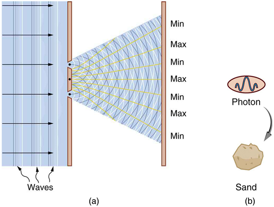
Electrons, once thought to be purely particles, also behave like waves. This concept, called wave-particle duality, was confirmed by the Davisson-Germer experiment, which showed electrons diffracting like waves when fired at a crystal. The de Broglie wavelength equation connects a particle's wavelength to its momentum, and it applies to all matter, not just electrons. Understanding this relationship is central to quantum mechanics and explains why particle behavior changes dramatically at very small scales.
Wave-Particle Duality
Wave behavior of electrons
In 1927, Clinton Davisson and Lester Germer fired a beam of electrons at a nickel crystal and observed something unexpected: the electrons produced a diffraction pattern, similar to the interference patterns created by X-rays (which were already known to be waves). This was direct evidence that electrons have wave-like properties.
The wavelength of those electrons matched a prediction made earlier by Louis de Broglie. He hypothesized that all matter exhibits wave-like behavior, with a wavelength given by:
- = wavelength of the particle
- = Planck's constant
- = momentum of the particle
De Broglie proposed this idea in 1924, three years before Davisson and Germer confirmed it experimentally. The key insight is that wave behavior isn't unique to light or electrons. Every object with momentum has an associated wavelength. The reason you don't notice your own wavelength is that for everyday objects, the wavelength is unimaginably small.
De Broglie wavelength calculations
Since momentum is the product of mass and velocity (), you can expand the de Broglie equation to:
where J·s (Planck's constant).
The wavelength is inversely proportional to momentum. That means:
- Higher momentum → shorter wavelength. A fast electron in a particle accelerator has a tiny wavelength.
- Lower momentum → longer wavelength. A slow electron in a low-energy microscope has a comparatively longer wavelength.
Wave effects like diffraction and interference only become noticeable when the de Broglie wavelength is comparable to the size of the objects the particle interacts with. For electrons, this typically means nanometer-scale structures like atoms or molecules. When the wavelength is close to the spacing between atoms in a crystal, electrons diffract and produce interference patterns. This is the basis of electron diffraction used in crystallography.
For a macroscopic object like a baseball (mass ~0.145 kg, speed ~40 m/s), the de Broglie wavelength works out to roughly m. That's far too small to ever detect, which is why wave behavior only matters at atomic and subatomic scales.
Quantum Mechanics and Wave Functions
Quantum mechanics is the theoretical framework that describes how matter and energy behave at atomic and subatomic scales. Within this framework, every particle is described by a wave function, a mathematical expression that encodes information about the particle's quantum state.
The wave function doesn't tell you exactly where a particle is. Instead, it gives you a probability amplitude. To find the actual probability of locating a particle at a particular position, you take the square of the wave function's magnitude. Regions where the squared magnitude is large are where you're most likely to find the particle; regions where it's small are where you're least likely to find it.
This probabilistic description is a direct consequence of matter's wave nature. Just as a water wave is spread out over space rather than concentrated at a single point, a particle's wave function is spread out, and the particle doesn't have a single definite position until a measurement is made.
Electron Microscopy
Because electrons have wave-like properties, they can be used to "see" things that visible light cannot. The resolution of any microscope is limited by the wavelength it uses. Visible light has wavelengths around 400–700 nm, so light microscopes can't resolve features much smaller than that. Electrons accelerated to high speeds have wavelengths on the order of picometers (thousandths of a nanometer), giving electron microscopes far better resolution.
Transmission vs scanning electron microscopes
Transmission Electron Microscope (TEM)
- A beam of electrons passes through a very thin sample (typically less than 100 nm thick).
- The electrons are diffracted by the sample's internal structure, and the resulting pattern forms an image on a detector or fluorescent screen.
- TEMs can achieve resolution down to about 0.1 nm, which is enough to image individual atoms.
- They reveal internal features like crystal lattice arrangements and structural defects.
- The tradeoff is that samples require extensive preparation, including slicing into ultra-thin sections and sometimes staining with heavy metals.
Scanning Electron Microscope (SEM)
- A focused beam of electrons scans across the surface of a sample point by point.
- The beam interacts with the surface, producing signals (secondary electrons, backscattered electrons, X-rays) that detectors collect to build an image.
- SEMs produce detailed 3D-looking images of surface features like roughness and texture.
- They can reach magnifications up to about 1,000,000× with resolution around 1 nm.
- Sample preparation is simpler than for TEM, and thicker, bulk samples can be imaged directly.
- By analyzing the X-rays emitted during scanning, SEMs can also identify the elemental composition of a sample (a technique called elemental mapping).
TEM vs. SEM in short: TEM shoots electrons through a thin sample to reveal internal structure at atomic resolution. SEM scans electrons across a surface to map topography and composition. TEM gives finer detail; SEM is more versatile with sample types.