🔋College Physics I – Introduction Unit 20 Review
20.7 Nerve Conduction–Electrocardiograms
20.7 Nerve Conduction–Electrocardiograms
Unit & Topic Study Guides
The Nature of Science and Physics
Kinematics
Two–Dimensional Kinematics
Force and Newton's Laws of Motion
Newton's Laws: Friction, Drag, and Elasticity
Circular Motion and Gravity
Work and Energy in Physics
Linear Momentum and Collisions
Statics and Torque
Rotational Motion & Angular Momentum
Fluid Statics
Fluid Dynamics: Biological & Medical Uses
Temperature and Gas Laws
Heat and Heat Transfer Methods
Thermodynamics
Oscillatory Motion and Waves
Physics of Hearing
Electric Charge and Fields
Electric Potential & Field
Electric Current, Resistance, and Ohm's Law
Circuits and DC Instruments
Magnetism
Electromagnetic Induction & AC Circuits
Electromagnetic Waves
Geometric Optics
Vision and Optical Instruments
Wave Optics
Special Relativity
Quantum Physics
Atomic Physics
Radioactivity and Nuclear Physics
Nuclear Physics in Medicine
Particle Physics
Nerve Conduction and Electrocardiograms
Nerve conduction and electrocardiograms (ECGs) are real-world applications of the electrical concepts covered in this unit. Neurons use differences in electric potential across their membranes to send signals, and the heart generates measurable voltage changes that doctors record as an ECG. Both topics connect current, resistance, and voltage to biological systems.
Neuron Signal Transmission
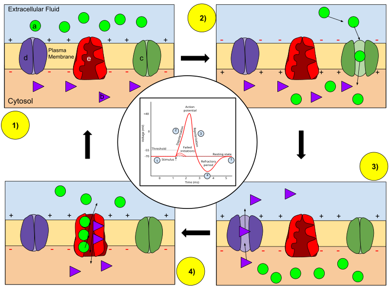
Neurons communicate using rapid changes in the voltage across their cell membranes. At rest, a neuron maintains a resting membrane potential of about , meaning the inside of the cell is negative relative to the outside. This voltage exists because of unequal concentrations of ions (mainly sodium and potassium ) on either side of the membrane, maintained by the sodium-potassium pump.
An action potential is the rapid voltage spike a neuron uses to send a signal. Here's how it unfolds:
- A stimulus raises the membrane potential to the threshold (around ).
- Voltage-gated channels open, and sodium rushes into the cell. The membrane potential shoots up toward . This phase is called depolarization.
- The channels close, and voltage-gated channels open. Potassium flows out of the cell, bringing the voltage back down. This is repolarization.
- The membrane briefly overshoots the resting potential (hyperpolarization) before the sodium-potassium pump restores the original .
After firing, the neuron enters a brief refractory period during which it cannot fire again. This prevents the signal from traveling backward.
The action potential propagates along the axon because the local current from one depolarized region triggers voltage-gated channels in the adjacent region, creating a wave-like signal from the cell body to the axon terminal. Sensory neurons carry these signals from receptors (touch, pain) to the brain, while motor neurons carry signals from the brain to muscles.
Role of Myelin Sheaths
Not all axons conduct signals at the same speed. Myelin sheaths, insulating layers of lipid and protein formed by glial cells, dramatically increase conduction velocity.
Instead of regenerating the action potential at every point along the axon, a myelinated neuron uses saltatory conduction: the signal "jumps" between nodes of Ranvier, small gaps in the myelin where voltage-gated ion channels are concentrated. This is faster and more energy-efficient because fewer ions need to be pumped back across the membrane.
- In the peripheral nervous system, Schwann cells wrap around axons to form myelin.
- In the central nervous system, oligodendrocytes serve the same role.
Myelination is especially important for long-distance signals, like those traveling from your spinal cord to your feet. Damage to myelin sheaths disrupts conduction and can cause serious neurological problems, as seen in multiple sclerosis.
Synaptic Transmission
When an action potential reaches the end of an axon (the axon terminal), the electrical signal has to cross a gap called the synaptic cleft to reach the next cell. This crossing works through a chemical process:
- The arriving action potential causes voltage-gated calcium channels to open at the axon terminal.
- Calcium influx triggers the release of neurotransmitter molecules into the synaptic cleft.
- Neurotransmitters diffuse across the cleft and bind to receptors on the target cell.
- This binding opens ion channels in the target cell, converting the chemical signal back into an electrical one.
So the sequence is: electrical → chemical → electrical. This mechanism allows neurons to communicate with other neurons, muscle cells, or glands.
Components of an ECG
An electrocardiogram (ECG) records the heart's electrical activity by measuring voltage differences between electrodes placed on the skin. The heart generates its own electrical signals, starting at the sinoatrial (SA) node in the right atrium, which acts as the natural pacemaker.
A normal ECG trace has three main features:
- P wave: Represents atrial depolarization. The atria contract and push blood into the ventricles.
- QRS complex: Represents ventricular depolarization. The ventricles contract and pump blood to the lungs and body. This is the largest feature because the ventricles have much more muscle mass than the atria. (Atrial repolarization happens at the same time but is hidden by the larger QRS signal.)
- T wave: Represents ventricular repolarization. The ventricles relax and begin to refill with blood.
Two important time intervals on the ECG:
- PR interval: Time from the start of atrial depolarization to the start of ventricular depolarization. It reflects how long the signal takes to travel through the AV node. A normal PR interval is about to .
- QT interval: Time from the start of ventricular depolarization to the end of ventricular repolarization. It reflects the total duration of ventricular electrical activity.
Abnormalities in the ECG waveform help doctors diagnose cardiac conditions:
- Arrhythmias such as atrial fibrillation (irregular P waves) or ventricular tachycardia (rapid QRS complexes)
- Conduction disorders like heart block (prolonged PR interval)
- Myocardial infarction (heart attack), often indicated by ST-segment elevation
- Electrolyte imbalances such as hypokalemia or hypercalcemia, which alter wave shapes and intervals