transforms the neural plate into the neural tube, forming the central nervous system. This process involves complex cell shape changes, signaling gradients, and precise closure mechanisms. Failed closure can lead to serious birth defects.

The plays a crucial role in nervous system patterning. It secretes , creating signaling gradients that establish distinct progenitor domains along the neural tube. These domains give rise to different types of neurons.

Neurulation and Neural Tube Formation

Neural Plate Formation and Folding

Top images from around the web for Neural Plate Formation and Folding
  • Neurulation transforms neural plate into neural tube forming central nervous system precursor
  • Neural plate induced by notochord signals along dorsal midline during
  • Neural plate folding driven by cell shape changes
    • Apical constriction causes cells to become wedge-shaped
    • Results in formation of neural groove
  • Neural folds elevate and converge towards midline
    • Eventually fuse to form neural tube in primary neurulation

Neural Tube Closure and Defects

  • Closure begins at multiple initiation sites
    • Proceeds in zipper-like fashion along embryo
  • Anterior and posterior neuropores close last
  • Secondary neurulation occurs in caudal region of some vertebrates
    • Neural tube forms through cavitation of solid cell cord
  • Failed closure leads to neural tube defects
    • (brain and skull do not develop)
    • (incomplete closure of spinal cord)

Notochord Signaling in Nervous System Patterning

Notochord as Primary Organizer

  • Rod-like mesodermal structure forms during gastrulation
  • Serves as primary organizer for developing nervous system
  • Secretes Sonic hedgehog (Shh) morphogen
    • Induces floor plate formation in ventral neural tube
  • Floor plate becomes secondary signaling center
    • Also secretes Shh establishing ventral-to-dorsal gradient

Signaling Gradients and Neural Tube Patterning

  • Bone Morphogenetic Proteins (BMPs) from dorsal antagonize Shh
  • Shh and BMP gradients create dorsal-ventral gene expression axis
  • Establishes distinct progenitor domains along neural tube
    • Ventral domains give rise to motor neurons
    • Dorsal domains produce sensory neurons
  • Notochord induces sclerotome formation in adjacent somites
    • Contributes to vertebral column development

Neural Crest Cell Formation and Differentiation

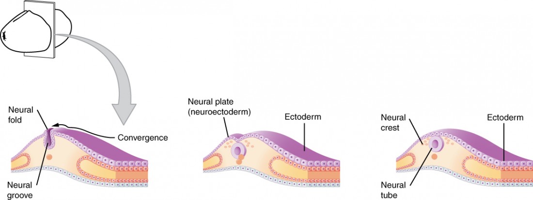
Neural Crest Induction and Migration

  • Multipotent cell population arises at neural plate and non-neural ectoderm border
  • Induction involves complex BMP, Wnt, and FGF signaling interplay
  • Undergo (EMT)
    • Delaminate from dorsal neural tube
    • Migrate extensively throughout embryo
  • Migration guided by intrinsic factors and environmental cues
    • Chemotactic signals (CXCL12, semaphorins)
    • Extracellular matrix interactions (fibronectin, laminin)

Neural Crest Derivatives and Contributions

  • Cranial forms craniofacial structures
    • Bones (maxilla, mandible)
    • Cartilage (nasal, ear)
    • Connective tissues of face and neck
  • Trunk neural crest gives rise to various cell types
    • Melanocytes in skin
    • Schwann cells of peripheral nervous system
    • Sympathetic and parasympathetic ganglia
  • Cardiac neural crest contributes to heart development
    • Forms outflow tract
    • Crucial for cardiac septation (dividing heart chambers)

Early Organogenesis and Primitive Gut Development

Primitive Gut Tube Formation

  • Organogenesis begins fourth week of human embryonic development
  • Primitive gut tube forms from
    • Initially straight tube divided into three regions
  • Foregut develops into:
    • Pharynx, esophagus, stomach, proximal duodenum
    • Associated organs (liver, pancreas)
  • Midgut forms:
    • Distal duodenum, jejunum, ileum
    • Cecum, appendix, ascending colon
    • Proximal two-thirds of transverse colon
  • Hindgut gives rise to:
    • Distal one-third of transverse colon
    • Descending colon, sigmoid colon, rectum
    • Upper part of anal canal

Gut Tube Patterning and Organ Specification

  • Differential growth rates cause gut tube folding
    • Forms specific organs
    • Positions organs within body cavity
  • Reciprocal interactions crucial for proper development
    • Endoderm and surrounding communicate
    • Guides organ specification (pancreatic buds, liver diverticulum)
  • Molecular signaling pathways direct regional identity
    • Sonic hedgehog (Shh) patterns foregut
    • Fibroblast growth factors (FGFs) influence hindgut development

Key Terms to Review (18)

Anencephaly: Anencephaly is a severe congenital condition that results from incomplete closure of the neural tube during embryonic development, leading to the absence of major parts of the brain and skull. This condition occurs when the anterior portion of the neural tube fails to close properly, which disrupts normal brain formation and regionalization, ultimately affecting the central nervous system's function. As a result, anencephaly is often classified as a type of neural tube defect and is associated with significant implications for early organogenesis and congenital disorders.
Anterior-posterior axis: The anterior-posterior axis is an essential body axis that defines the orientation of an organism from the head (anterior) to the tail (posterior). This axis plays a crucial role in establishing the overall body plan and organization during development, influencing various processes such as segmentation, organ positioning, and overall morphological patterning.
Bmp signaling: BMP signaling refers to the Bone Morphogenetic Protein signaling pathway, which is crucial in various developmental processes, including cell differentiation, migration, and organ formation. This pathway plays a significant role in the development and patterning of tissues, influencing key events such as body axis establishment and germ layer formation.
Dorsal-ventral axis: The dorsal-ventral axis is a fundamental body axis that defines the back (dorsal) and belly (ventral) orientation of an organism. This axis is crucial for the proper spatial organization of body structures and plays a key role in developmental processes, including the establishment of body plans and the formation of various organ systems.
Ectoderm: Ectoderm is one of the three primary germ layers formed during embryonic development, specifically the outermost layer that gives rise to various structures in the body. This layer plays a critical role in the development of the nervous system, skin, and several other organs. Understanding ectoderm is essential for comprehending how complex structures arise from simple embryonic layers during processes like neurulation, organogenesis, gastrulation, and blastulation.
Endoderm: The endoderm is one of the three primary germ layers formed during embryonic development, specifically giving rise to the innermost layers of tissues and organs in an organism. It plays a crucial role in forming the lining of the digestive tract and respiratory systems, and it is responsible for generating many internal organs, such as the liver and pancreas, which are essential for bodily functions.
Epithelial-to-Mesenchymal Transition: Epithelial-to-mesenchymal transition (EMT) is a biological process where epithelial cells lose their characteristics, such as cell-cell adhesion and polarity, and gain mesenchymal properties, which include increased motility and invasiveness. This transformation is crucial during various developmental processes, such as cell migration during neural crest formation, the early stages of organ development, and the formation of germ layers. EMT allows cells to adapt to new environments, facilitating important transitions in tissue architecture and function.
Gastrulation: Gastrulation is a fundamental phase in embryonic development where the single-layered blastula reorganizes into a multi-layered structure called the gastrula, forming the three primary germ layers: ectoderm, mesoderm, and endoderm. This process sets the stage for the development of various tissues and organs in the body and plays a crucial role in establishing the body axes and overall architecture of the organism.
Mesoderm: Mesoderm is one of the three primary germ layers formed during embryonic development, lying between the ectoderm and endoderm. This layer gives rise to various structures, including muscles, bones, the circulatory system, and the excretory system, playing a crucial role in organ development and body plan organization.
Neural Crest: Neural crest refers to a unique population of cells that arise during the development of vertebrates, specifically from the border between the neural tube and the ectoderm. These cells are multipotent, meaning they can differentiate into various cell types and contribute to the formation of diverse structures, including the peripheral nervous system, facial cartilage, and melanocytes. The emergence of neural crest cells is crucial for many developmental processes and connects closely to the events that occur during neurulation and early organogenesis, as well as the gastrulation phase when germ layers are formed.
Neural tube formation: Neural tube formation is a critical developmental process where the neural plate folds to create the neural tube, which ultimately gives rise to the central nervous system, including the brain and spinal cord. This process is crucial for proper organogenesis and involves precise cellular movements, signaling pathways, and the coordination of various embryonic tissues.
Neuroectoderm: Neuroectoderm is a specialized region of the ectoderm that gives rise to the nervous system, including the brain and spinal cord, during embryonic development. It plays a crucial role in neurulation, where the neural plate forms and folds to create the neural tube, setting the stage for early organogenesis and the establishment of neural structures.
Neurulation: Neurulation is the developmental process during which the neural plate forms and folds to create the neural tube, which eventually develops into the central nervous system. This process is critical for establishing the organization of the brain and spinal cord and is influenced by various signaling pathways that dictate the fate of neural progenitor cells, setting the stage for further organogenesis and the evolutionary context of vertebrate development.
Notochord: The notochord is a flexible rod-like structure that provides support and defines the primitive axis of the developing embryo. It plays a crucial role during early development by serving as a key signaling center that influences the formation of surrounding structures, particularly in the development of the nervous system and vertebral column.
Somitogenesis: Somitogenesis is the process by which somites, the segmented blocks of mesodermal tissue, form along the developing neural tube in an embryo. This critical process occurs during early embryonic development and plays a key role in organizing the body plan by giving rise to structures such as vertebrae, skeletal muscles, and dermis. Somitogenesis is intricately linked to other developmental processes, including neurulation and organogenesis, as it helps define the segmentation of the body axis.
Sonic Hedgehog: Sonic Hedgehog is a signaling protein that plays a crucial role in embryonic development, particularly in the regulation of cell growth, differentiation, and tissue patterning. This protein is essential for the formation of various structures in the body, including limbs, brain, and organs, and its signaling pathway is integral to establishing body axes and ensuring proper organ development.
Spina bifida: Spina bifida is a congenital defect that occurs when the neural tube, which eventually develops into the spine and surrounding structures, fails to close completely during early embryonic development. This condition results in varying degrees of damage to the spinal cord and nerves, leading to a range of physical and neurological impairments. The severity of spina bifida can vary, impacting motor function, sensory perception, and even bladder or bowel control.
Wnt Signaling: Wnt signaling is a complex network of proteins that play crucial roles in regulating cellular processes such as cell proliferation, differentiation, and migration during development. This pathway is integral for establishing body axes, forming germ layers, and guiding various developmental events, including organogenesis and tissue regeneration.
© 2024 Fiveable Inc. All rights reserved.
AP® and SAT® are trademarks registered by the College Board, which is not affiliated with, and does not endorse this website.