Proteins are the workhorses of life, and their structure is key to their function. From simple chains of amino acids to complex 3D shapes, proteins fold into specific structures that determine how they work in our bodies.
Understanding protein structure is crucial for grasping how they function. We'll look at the different levels of protein structure, from primary to quaternary, and see how each level builds on the last to create functional molecules.
Protein Structure and Function
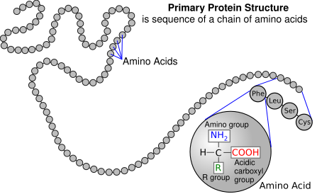
Primary Structure and Higher-Order Structures
- The primary structure of a protein is the linear sequence of amino acids connected by peptide bonds, which are covalent bonds formed between the carboxyl group of one amino acid and the amino group of the next
- The primary structure is determined by the genetic code, which specifies the order of amino acids in a protein
- The sequence of amino acids in the primary structure determines the higher-order structures (secondary, tertiary, and quaternary) of the protein as the chemical properties of the amino acids influence the interactions and folding of the polypeptide chain
- The primary structure also determines the protein's unique function as the specific sequence of amino acids defines the protein's shape, binding sites, and catalytic properties
- For example, the primary structure of hemoglobin dictates its ability to bind and transport oxygen in the blood
- Enzymes, such as trypsin, have specific active sites determined by their primary structure that allow them to catalyze reactions
Secondary Structures of Proteins
Alpha Helices and Beta Sheets
- Secondary structures are the local, regular arrangements of amino acids within a polypeptide chain, primarily stabilized by hydrogen bonds between the main-chain peptide groups
- The two most common secondary structures are alpha helices and beta sheets
- Alpha helices are right-handed spiral conformations, with the main-chain carbonyl oxygen of each amino acid forming a hydrogen bond with the main-chain amino hydrogen of the amino acid four residues ahead in the sequence
- For example, the alpha helix is a common motif in the keratin proteins that make up hair and nails
- Beta sheets are extended conformations with two or more beta strands arranged either parallel or antiparallel to each other, forming hydrogen bonds between the main-chain peptide groups of adjacent strands
- An example of a protein with beta sheets is the immunoglobulin G (IgG) antibody, which has a beta-sandwich structure in its constant regions

Factors Influencing Secondary Structure Formation
- The formation of secondary structures is influenced by the chemical properties of the amino acids in the primary sequence, such as their hydrophobicity, charge, and size
- Secondary structures contribute to the overall stability and folding of the protein by reducing the exposure of hydrophobic amino acids to the aqueous environment and maximizing the formation of hydrogen bonds
- Proline and glycine are often found in turns or loops between secondary structures due to their unique conformational properties
- Amino acids with hydrophobic side chains (leucine, valine, isoleucine) are more likely to be found in the interior of alpha helices or beta sheets
Tertiary Structure and Stability
Three-Dimensional Arrangement and Non-Covalent Interactions
- Tertiary structure refers to the three-dimensional arrangement of all amino acids in a polypeptide chain, resulting from the folding and spatial organization of secondary structures
- The tertiary structure is stabilized by various non-covalent interactions, including hydrogen bonds, hydrophobic interactions, van der Waals forces, and ionic interactions (salt bridges) between amino acid side chains
- For example, the tertiary structure of myoglobin, a protein that stores oxygen in muscle cells, is stabilized by extensive hydrophobic interactions between the non-polar amino acids in its interior
- Disulfide bonds, which are covalent bonds formed between the thiol groups of cysteine residues, can also contribute to the stability of the tertiary structure
- Insulin, a hormone that regulates blood sugar levels, contains two polypeptide chains linked by disulfide bonds that are crucial for its tertiary structure and function
Hydrophobic Effect and Protein Function
- The hydrophobic effect plays a crucial role in the folding and stability of the tertiary structure, as hydrophobic amino acids tend to cluster in the protein's interior, away from the aqueous environment
- The tertiary structure of a protein determines its unique shape, which is essential for its biological function, such as ligand binding, catalysis, or interaction with other molecules
- The tertiary structure of enzymes creates specific active sites that bind substrates and catalyze reactions
- Membrane proteins, such as ion channels and receptors, have tertiary structures that allow them to span the lipid bilayer and perform their functions
- Factors that disrupt the tertiary structure, such as changes in temperature, pH, or the presence of denaturants, can lead to protein unfolding and loss of function
- Heat and urea are common denaturants that disrupt the non-covalent interactions stabilizing the tertiary structure, causing the protein to unfold and lose its native conformation
Quaternary Structure and Function
Multi-Subunit Protein Complexes
- Quaternary structure refers to the arrangement of two or more polypeptide chains (subunits) that associate to form a multi-subunit protein complex
- Proteins with quaternary structure are called oligomers or multimers, and the individual polypeptide chains are called protomers or subunits
- Hemoglobin, the oxygen-carrying protein in red blood cells, is a tetramer composed of two alpha and two beta subunits
- Glutamate dehydrogenase, an enzyme involved in amino acid metabolism, is a homohexamer made up of six identical subunits
- The association of subunits in a quaternary structure is stabilized by the same non-covalent interactions that contribute to the tertiary structure, such as hydrogen bonds, hydrophobic interactions, van der Waals forces, and ionic interactions
- The subunits in a quaternary structure can be identical (homooligomers) or different (heterooligomers), and their arrangement can be symmetric or asymmetric
Functional Significance and Regulation
- Quaternary structure allows for the formation of large, complex proteins with multiple functional domains, enabling them to carry out sophisticated biological processes, such as oxygen transport (hemoglobin) or enzyme regulation (allosteric enzymes)
- The quaternary structure of hemoglobin allows for cooperative binding of oxygen, enhancing its ability to transport oxygen in the blood
- Allosteric enzymes, such as aspartate transcarbamoylase, have regulatory sites distant from the active site that modulate enzyme activity through conformational changes in the quaternary structure
- The assembly and disassembly of subunits in a quaternary structure can be regulated by various factors, such as ligand binding, post-translational modifications, or changes in the cellular environment, providing an additional level of control over protein function
- The binding of 2,3-bisphosphoglycerate (2,3-BPG) to hemoglobin modulates its affinity for oxygen, facilitating oxygen release in tissues
- The phosphorylation of glycogen phosphorylase by phosphorylase kinase triggers a conformational change in its quaternary structure, activating the enzyme and promoting glycogen breakdown