Emerging techniques in biophysical chemistry are revolutionizing how we study biological systems. , , , and offer new ways to visualize and analyze macromolecules, overcoming limitations of traditional methods.

These advances enable researchers to examine , , and interactions with incredible detail. By combining these techniques, scientists can gain deeper insights into complex biological processes, pushing the boundaries of our understanding in biophysical chemistry.

Emerging Techniques in Biophysical Chemistry

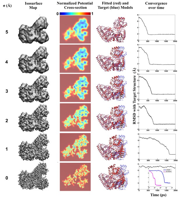
Cryo-Electron Microscopy (Cryo-EM)

  • Rapidly freezes biological samples in liquid ethane
  • Images samples using electron microscopy
  • Visualizes macromolecular structures in their native state without the need for crystallization or staining

Super-Resolution Microscopy

  • Encompasses techniques such as stimulated emission depletion (STED) microscopy, (SIM), and ()
  • Allows for imaging beyond the diffraction limit of light
  • Enables visualization of biological structures at nanometer-scale resolution

Other Emerging Techniques

  • (AFM) provides high-resolution topographical imaging and force measurements of biological samples
  • (SAXS) allows for the study of macromolecular structures in solution

Cryo-EM vs Traditional Microscopy

Advantages of Cryo-EM

  • Enables visualization of large and cellular structures in their native state
  • Avoids potential artifacts introduced by sample preparation methods required for traditional electron microscopy (fixation and staining)

Advantages of Super-Resolution Microscopy

  • Overcomes the diffraction limit of light
  • Allows for visualization of biological structures at resolutions previously only achievable by electron microscopy
  • Enables the study of dynamic processes and in living cells

Advantages of Other Emerging Techniques

  • AFM provides high-resolution topographical imaging of biological samples in their native environment without requiring sample fixation or staining
  • SAXS provides information on the size, shape, and flexibility of biological molecules in solution without the need for crystallization

Applications of Emerging Techniques

Protein Structure and Dynamics

  • Cryo-EM has been instrumental in determining of large protein complexes (ribosomes, ion channels, and viral capsids)
  • Cryo-EM has enabled visualization of proteins in different conformational states, providing insights into their
  • Super-resolution microscopy techniques have been applied to study the organization and dynamics of protein complexes in living cells (clustering of receptors in the plasma membrane and assembly of cytoskeletal structures)

Protein Interactions and Mechanical Properties

  • AFM has been used to study the of proteins (unfolding and refolding of individual protein molecules under force)
  • AFM has been applied to investigate protein-protein and at the single-molecule level
  • SAXS has been employed to study of proteins in solution (folding and unfolding of proteins in response to changes in temperature or pH)
  • SAXS has been used to characterize interactions between proteins and other macromolecules (nucleic acids and lipids)

Impact on Biophysical Chemistry Research

Advancements in Cryo-EM

  • Increasing resolution and sensitivity of cryo-EM are expected to enable the determination of structures for a wider range of protein complexes and cellular components
  • Provides new insights into the molecular basis of biological processes

Developments in Super-Resolution Microscopy

  • Continued development of super-resolution microscopy techniques is likely to enhance our understanding of the spatial organization and dynamics of biomolecules in living cells
  • Leads to new discoveries in cell biology and molecular biophysics

Progress in AFM and SAXS

  • Advances in AFM technology (high-speed imaging and multiparametric measurements) are expected to expand its applications in studying the mechanical properties and interactions of biological systems at the nanoscale level
  • Integration of SAXS with other biophysical techniques (nuclear magnetic resonance (NMR) spectroscopy and ) is likely to provide a more comprehensive understanding of the structure, dynamics, and interactions of biomolecules in solution

Combination of Emerging Techniques

  • The combination of these emerging techniques with other approaches ( and ) is expected to enable the study of complex biological systems with unprecedented detail and resolution
  • Leads to new breakthroughs in biophysical chemistry research

Key Terms to Review (26)

Afm: Atomic force microscopy (AFM) is a powerful imaging technique used to visualize surfaces at the nanoscale by measuring the interaction forces between a sharp probe and the sample surface. AFM allows researchers to obtain high-resolution images of materials and biological samples, making it essential for studying single molecules and their interactions, as well as contributing to the development of new biophysical techniques.
Atomic Force Microscopy: Atomic Force Microscopy (AFM) is a powerful imaging technique that enables the visualization and manipulation of surfaces at the atomic level using a sharp tip attached to a cantilever. This technique allows researchers to study materials, biological samples, and nanoscale structures with high resolution, contributing significantly to understanding various molecular interactions and topological features.
Conformational Changes: Conformational changes refer to the alterations in the three-dimensional shape of a molecule, often as a response to environmental factors or interactions with other molecules. These changes can significantly impact the functionality of biomolecules, affecting processes such as enzyme activity, molecular recognition, and motor protein movement. Understanding these changes is crucial for insights into molecular dynamics and mechanisms underlying biological functions.
Cryo-electron microscopy: Cryo-electron microscopy (cryo-EM) is an advanced imaging technique used to visualize biological samples at near-atomic resolution while preserving their native state by freezing them in liquid ethane or propane. This method allows scientists to study the structure of macromolecules and complexes in their functional forms, bridging the gap between traditional electron microscopy and X-ray crystallography.
Cryo-EM: Cryo-electron microscopy (cryo-EM) is a powerful imaging technique that allows researchers to visualize biological macromolecules and complexes at near-atomic resolution while preserving their native structures in a frozen state. This method has revolutionized structural biology by enabling the study of proteins, viruses, and other biomolecules without the need for crystallization or staining, thus maintaining their functional conformation.
Dynamics: Dynamics refers to the study of forces and their effects on the behavior of physical systems over time. This concept is crucial in understanding how different components within a system interact and change, which is fundamental to analyzing complex biological networks and the mechanisms driving molecular interactions in biophysical chemistry. By examining the dynamics, researchers can gain insights into the stability, fluctuations, and adaptability of various systems.
Functional dynamics: Functional dynamics refers to the study of the motions and interactions of biomolecules and their functions in biological systems. This concept focuses on how the structural changes in biomolecules, such as proteins and nucleic acids, influence their biological activities and roles within cells. Understanding functional dynamics is essential for revealing how molecular flexibility, conformational changes, and dynamic interactions contribute to various biochemical processes.
High-resolution structures: High-resolution structures refer to detailed three-dimensional arrangements of biological macromolecules, such as proteins and nucleic acids, determined using advanced techniques that provide atomic-level clarity. These structures are crucial for understanding the precise interactions and functions of biomolecules, significantly enhancing our knowledge of biochemical processes and molecular mechanisms.
Macromolecular complexes: Macromolecular complexes are large, often multi-component assemblies formed by the interaction of various macromolecules such as proteins, nucleic acids, and carbohydrates. These complexes play crucial roles in cellular processes, serving as the machinery for various biological functions including enzymatic reactions, signal transduction, and structural support. Their formation is influenced by factors like molecular interactions, structural conformations, and environmental conditions, which can be analyzed through emerging techniques in biophysical chemistry.
Mass Spectrometry: Mass spectrometry is an analytical technique used to measure the mass-to-charge ratio of ions, allowing for the identification and quantification of various compounds in a sample. This powerful method is widely used in biochemistry and biophysical chemistry for analyzing biomolecules, understanding complex mixtures, and determining molecular structures. The information obtained from mass spectrometry plays a crucial role in areas such as drug development, proteomics, and lipid analysis, highlighting its interdisciplinary nature and applications.
Mechanical Properties: Mechanical properties refer to the physical characteristics of materials that describe their behavior when subjected to mechanical forces. These properties include elasticity, tensile strength, and viscosity, which play a critical role in understanding how biological molecules respond to various stresses and strains, particularly in the context of emerging techniques in biophysical chemistry that aim to probe these characteristics at molecular levels.
Molecular dynamics simulations: Molecular dynamics simulations are computational methods used to model the behavior of molecular systems over time by solving Newton's equations of motion. These simulations provide insights into the dynamic behavior of biomolecules, allowing researchers to explore their structure, dynamics, and interactions in a way that complements experimental techniques. This approach is fundamental in understanding the physical principles governing molecular behavior and is increasingly employed in new technologies within biophysical chemistry.
Molecular interactions: Molecular interactions refer to the various attractive and repulsive forces that occur between molecules, influencing their behavior, structure, and function. These interactions play a crucial role in determining the properties of biological systems, affecting how molecules bind to one another, how they aggregate, and how they perform their functions within cells. Understanding these interactions is fundamental to biophysical chemistry, especially when analyzing single molecules and developing new experimental techniques.
NMR Spectroscopy: NMR (Nuclear Magnetic Resonance) spectroscopy is a powerful analytical technique used to determine the structure and dynamics of molecules by measuring the magnetic properties of atomic nuclei. This method provides insights into molecular environments and interactions, making it essential in studying biomolecules, including proteins and nucleic acids.
Nuclear magnetic resonance spectroscopy: Nuclear magnetic resonance spectroscopy (NMR) is a powerful analytical technique used to determine the structure and dynamics of molecules by observing the interaction of atomic nuclei with an external magnetic field. This method provides insight into molecular environments and interactions, making it invaluable in the study of biomolecules, such as proteins and nucleic acids, within biophysical chemistry.
Protein Structures: Protein structures refer to the specific three-dimensional arrangements of amino acids that make up proteins, which are essential macromolecules in biological systems. Understanding these structures is crucial as they dictate the function of proteins, and advanced techniques in biophysical chemistry allow for detailed visualization and analysis of these conformations, enabling insights into their roles in biological processes.
Protein-ligand interactions: Protein-ligand interactions refer to the specific binding events between a protein and a small molecule (ligand) that can trigger biological responses or influence protein activity. These interactions are fundamental in various biochemical processes, impacting areas like enzyme activity, signal transduction, and molecular recognition. Understanding these interactions is essential for exploring binding equilibria and kinetics, employing techniques like NMR spectroscopy for structural analysis, and investigating new methods in biophysical chemistry.
Protein-protein interactions: Protein-protein interactions refer to the specific ways in which two or more proteins bind and interact with each other to form complexes that are crucial for various biological processes. These interactions can influence protein function, stability, localization, and can be involved in signal transduction pathways, enzyme activity regulation, and the formation of cellular structures. Understanding these interactions is essential for studying cellular functions and developing therapeutic strategies.
SAXS: Small Angle X-ray Scattering (SAXS) is a powerful analytical technique used to determine the structural properties of macromolecules in solution at low resolutions. SAXS measures the scattering of X-rays at small angles to provide information about the size, shape, and overall arrangement of particles, such as proteins and polymers, in a sample. This technique is particularly valuable for studying biological systems as it can be applied to samples in their native environments, allowing researchers to gather insights into molecular dynamics and interactions.
Single-molecule fluorescence spectroscopy: Single-molecule fluorescence spectroscopy is a powerful analytical technique that allows for the observation and measurement of individual molecules in a sample, providing insights into their dynamic behavior and interactions. This method enhances our understanding of molecular processes by measuring fluorescence emitted from a single molecule, enabling researchers to explore the heterogeneity and kinetics of complex biological systems with high spatial and temporal resolution.
Single-molecule localization microscopy: Single-molecule localization microscopy is a high-resolution imaging technique that allows researchers to visualize and track individual molecules within cells. This method overcomes the diffraction limit of conventional microscopy by utilizing the precise localization of fluorescently tagged molecules, enabling the study of dynamic biological processes at the nanoscale.
Small-angle x-ray scattering: Small-angle x-ray scattering (SAXS) is a powerful technique used to analyze the structure of materials at the nanoscale by measuring the scattering of x-rays off of particles in solution. This method provides valuable insights into the shape, size, and distribution of macromolecules and complexes, making it essential for studying biological systems and materials science.
Smlm: smlm, or single-molecule localization microscopy, is a cutting-edge imaging technique used to visualize and track individual molecules in biological samples with high spatial and temporal resolution. This method allows scientists to bypass the diffraction limit of light, enabling the observation of structures and dynamics at the nanoscale level, which is crucial for understanding complex biological processes.
Stimulated emission depletion microscopy: Stimulated emission depletion microscopy (STED) is a super-resolution imaging technique that enhances the resolution of fluorescence microscopy by using a combination of focused laser beams to deplete fluorescence in specific areas while allowing it to emit in others. This method enables scientists to visualize cellular structures at nanometer-scale resolution, which is crucial for understanding complex biological processes.
Structured illumination microscopy: Structured illumination microscopy (SIM) is an advanced imaging technique that enhances the resolution of fluorescence microscopy by using a patterned light source to excite the sample. This method allows for the capture of high-resolution images that surpass the diffraction limit of conventional light microscopy, making it invaluable for studying cellular structures and dynamics in biological samples.
Super-resolution microscopy: Super-resolution microscopy is a collection of advanced imaging techniques that surpass the diffraction limit of light to visualize biological structures at a much higher resolution than traditional optical microscopy. This innovative approach allows researchers to observe cellular components with unprecedented clarity, leading to deeper insights into biological processes and molecular interactions.
© 2024 Fiveable Inc. All rights reserved.
AP® and SAT® are trademarks registered by the College Board, which is not affiliated with, and does not endorse this website.