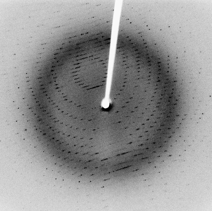
X-ray crystallography is a powerful technique for revealing the 3D structure of molecules. It uses X-ray diffraction patterns from crystals to map electron density, showing atom positions. This method is crucial for understanding protein structures and functions.

Protein crystallization is a key step, involving careful control of conditions to grow high-quality crystals. Once obtained, these crystals are analyzed to create detailed electron density maps, which are then used to build and refine molecular models.

X-ray Crystallography Principles

Fundamentals of X-ray Diffraction

Top images from around the web for Fundamentals of X-ray Diffraction
  • X-ray crystallography utilizes the diffraction of X-rays by the regularly spaced atoms in a crystal to determine the three-dimensional structure of molecules
  • The diffraction pattern produced when X-rays interact with a crystalline sample is recorded and analyzed to determine the electron density distribution within the crystal
  • Fourier transform is applied to the diffraction data to convert it into an , which reveals the positions of atoms in the molecule

Factors Affecting Structural Determination

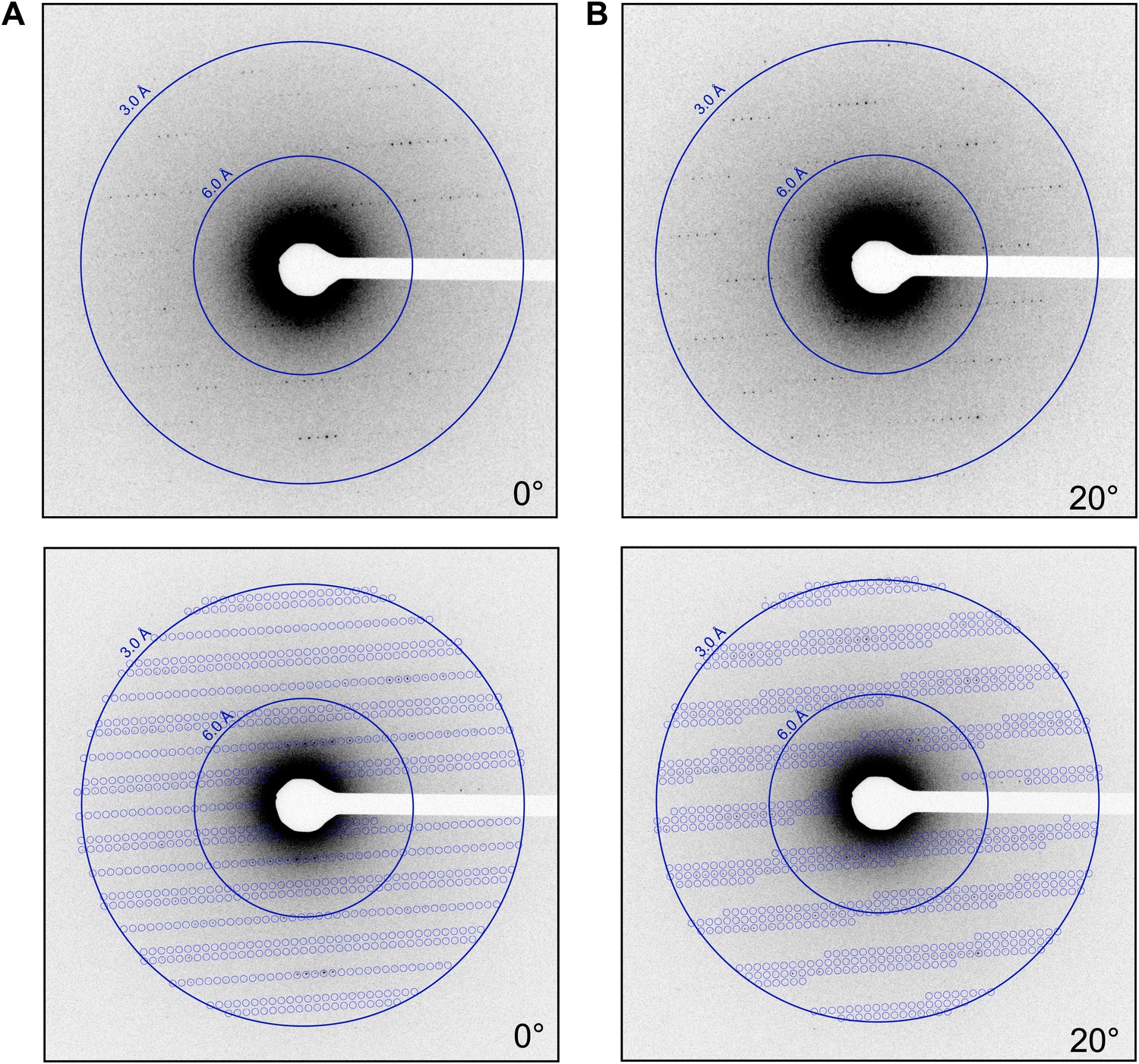
  • The quality and resolution of the electron density map depend on factors such as crystal size, purity, and the wavelength of the X-rays used
  • Molecular models are built by fitting the known amino acid sequence of the protein into the electron density map, followed by refinement to improve the agreement between the model and the experimental data
  • X-ray crystallography can determine the structure of proteins, nucleic acids, and other biological macromolecules at atomic resolution, typically in the range of 1-3 Å (angstroms)
  • The three-dimensional structures obtained by X-ray crystallography provide insights into the function, interactions, and mechanisms of action of biological molecules (enzymes, receptors)

Protein Crystallization Process

Methods and Techniques

  • Protein crystallization is the process of inducing the formation of ordered, repeating arrays of protein molecules in a supersaturated solution
  • The most common method for protein crystallization is vapor diffusion, which involves mixing the protein solution with a precipitant solution and allowing the mixture to equilibrate against a reservoir containing a higher concentration of the precipitant
  • As water vapor diffuses from the protein drop to the reservoir, the protein concentration increases, leading to supersaturation and potentially crystal and growth
  • Seeding techniques, such as microseeding or macroseeding, can be used to promote and improve crystal quality by introducing preformed crystal nuclei into the crystallization drop

Factors Influencing Crystallization Success

  • Factors that influence protein crystallization include protein purity, concentration, pH, temperature, and the presence of additives or ligands (metal ions, cofactors)
  • The choice of precipitant, such as salts (ammonium sulfate), polymers (polyethylene glycol), or organic solvents (isopropanol), can significantly impact the success of crystallization and the quality of the resulting crystals
  • The quality of protein crystals is assessed by their size, morphology, and the resolution of the diffraction pattern they produce when exposed to X-rays
  • High-quality crystals are essential for obtaining accurate and high-resolution structural information by X-ray crystallography

Electron Density Maps

Interpretation of Electron Density

  • Electron density maps represent the three-dimensional distribution of electrons in the crystal, which corresponds to the positions of atoms in the molecule
  • The electron density at a given point in the map is proportional to the number of electrons present at that location, with higher density indicating the presence of heavier atoms or multiple atoms
  • The resolution of the electron density map determines the level of detail visible, with higher resolution maps allowing for the identification of individual atoms and the precise positioning of side chains
  • The interpretation of electron density maps involves the recognition of characteristic patterns corresponding to specific amino acid residues (glycine, tryptophan), secondary structures (alpha helices, beta sheets), and other structural features of the molecule

Model Building and Refinement

  • Molecular model building is the process of fitting the known amino acid sequence of the protein into the electron density map, using the map as a guide for positioning the atoms
  • The initial model is typically built using automated or semi-automated methods, followed by manual adjustments to improve the fit to the electron density
  • Model refinement is an iterative process that involves adjusting the atomic positions, B-factors (atomic displacement parameters), and occupancies to minimize the difference between the calculated and observed diffraction data
  • Throughout the refinement process, the quality of the model is assessed using various validation tools and statistical measures, such as the and the Ramachandran plot, to ensure its accuracy and reliability

Applications of X-ray Crystallography

Drug Discovery and Design

  • X-ray crystallography plays a crucial role in drug discovery by providing detailed structural information on drug targets, such as enzymes (kinases), receptors (G protein-coupled receptors), and transport proteins (ion channels)
  • The three-dimensional structures of target proteins obtained by X-ray crystallography enable the identification of binding sites and the design of molecules that can specifically interact with these sites to modulate protein function
  • Structure-based drug design involves the use of protein-ligand complex structures to guide the optimization of lead compounds, improving their potency, selectivity, and pharmacokinetic properties

Protein Engineering and Structure-Function Relationships

  • In protein engineering, X-ray crystallography is used to guide the rational design of proteins with enhanced stability, specificity, or catalytic activity
  • By comparing the structures of wild-type and mutant proteins, researchers can gain insights into the molecular basis of protein function and the effects of specific mutations on protein stability and activity
  • X-ray crystallography is instrumental in elucidating the structure-function relationships of biomolecules, providing a detailed understanding of how the three-dimensional arrangement of atoms determines the biological properties of proteins, nucleic acids, and their complexes
  • Comparative structural analysis of related proteins from different organisms or protein families can reveal evolutionary relationships and provide insights into the molecular mechanisms of protein function and regulation (enzyme catalysis, signal transduction)
  • The integration of structural information obtained by X-ray crystallography with other experimental data, such as biochemical, biophysical, and computational studies, enables a comprehensive understanding of the structure, dynamics, and function of biological systems at the molecular level

Key Terms to Review (18)

Bragg's Law: Bragg's Law is a fundamental principle in X-ray crystallography that describes the relationship between the angle of incidence of X-rays and the distance between atomic planes in a crystal lattice. It states that constructive interference occurs when the path difference between X-rays reflected from adjacent planes is an integer multiple of the wavelength, enabling the determination of crystal structures.
Crystal Growth: Crystal growth is the process by which a solid crystalline structure forms from a solution, vapor, or melt as molecules or ions arrange themselves in a repeating pattern. This process is critical in determining the physical properties of materials, influencing everything from the quality of a crystal to its suitability for techniques like X-ray crystallography, which analyzes crystal structures to reveal important molecular information.
Detector: A detector is a device or component that measures and responds to physical signals or changes in an environment, converting them into readable data for analysis. In various scientific techniques, detectors are crucial as they enable the visualization and quantification of the information obtained from samples, making them essential for understanding molecular structures and interactions.
Electron density map: An electron density map is a three-dimensional representation of the distribution of electron density in a molecule, created from X-ray diffraction data. This map is crucial for visualizing the positions of atoms within a crystal structure and understanding molecular interactions. By interpreting the electron density, researchers can deduce the arrangement of atoms, identify functional groups, and analyze conformational changes in biomolecules.
James D. Watson: James D. Watson is an American molecular biologist, geneticist, and zoologist best known for co-discovering the double helix structure of DNA alongside Francis Crick. His work has had a profound impact on the field of genetics and molecular biology, particularly in the use of X-ray crystallography to reveal the structural complexities of DNA.
Ligand binding analysis: Ligand binding analysis is the study of the interaction between a ligand, which can be a small molecule or a protein, and its target, typically a receptor or an enzyme. This analysis is crucial for understanding the affinity and specificity of the ligand towards its target, which can inform drug design and biochemical pathways. It utilizes various experimental techniques to quantify binding events, revealing important information about molecular interactions and biological functions.
Molecular modeling: Molecular modeling is a computational technique used to represent and manipulate molecular structures to understand their properties and behaviors. This approach combines principles from chemistry, physics, and mathematics to create visual models that help in predicting molecular interactions and reactions. By utilizing molecular modeling, researchers can visualize three-dimensional structures, analyze molecular dynamics, and simulate various chemical processes, which are essential in fields such as drug design and materials science.
Nucleation: Nucleation is the initial process that occurs when small clusters or 'nuclei' form from a supersaturated solution or vapor, which can lead to the growth of larger structures such as crystals or aggregates. This process is critical in understanding how misfolded proteins aggregate and contribute to various diseases, as well as in the formation of crystalline structures analyzed through techniques like X-ray crystallography. Nucleation can be either homogeneous, occurring spontaneously, or heterogeneous, where pre-existing surfaces facilitate the process.
Phase problem: The phase problem refers to the challenge encountered in X-ray crystallography where the phase information of diffracted X-rays is lost during the measurement process. This loss of phase information complicates the reconstruction of the electron density map, which is essential for determining the three-dimensional structure of molecules. Overcoming the phase problem is crucial for accurate structural analysis and has led to the development of various computational techniques and methods.
Powder diffraction: Powder diffraction is a technique used to determine the crystalline structure of materials by analyzing the diffraction pattern produced when X-rays are scattered by a powdered sample. This method is particularly useful for identifying unknown substances and determining their atomic arrangements, as it provides information on the symmetry and dimensions of the unit cell, which are fundamental in crystallography.
Protein structure determination: Protein structure determination is the process of identifying the three-dimensional arrangement of atoms within a protein molecule. This involves techniques that provide insights into the protein's function, interactions, and stability, ultimately revealing how proteins work at a molecular level. Understanding protein structure is crucial for various applications, including drug design and understanding diseases caused by protein misfolding.
R-factor: The r-factor, or R-factor, is a critical metric used in X-ray crystallography to measure the quality of the data collected and the accuracy of the resulting model of a crystal structure. It indicates how well the observed diffraction data matches the calculated data from the model, with lower values suggesting a better fit and higher reliability of the structural information derived from X-ray analysis.
Reciprocal Lattice: A reciprocal lattice is a conceptual lattice used in crystallography that represents the periodicity of a crystal structure in momentum space, rather than real space. It is crucial for understanding diffraction patterns and the behavior of waves, such as X-rays or electrons, interacting with a crystalline material. The reciprocal lattice relates to the real lattice of the crystal by Fourier transformation, helping to analyze various properties such as electron behavior and crystal symmetries.
Refinement software: Refinement software is a type of computational tool used to enhance the accuracy and quality of molecular models obtained through techniques like X-ray crystallography. It systematically adjusts the positions of atoms in a crystal structure to minimize the difference between observed and calculated diffraction data, resulting in a more reliable and accurate representation of the molecule's three-dimensional structure.
Rosalind Franklin: Rosalind Franklin was a pioneering scientist whose X-ray diffraction images of DNA were critical in revealing the double helical structure of DNA. Her work laid the foundation for understanding the molecular structure of DNA, which is essential in genetics and biophysics, connecting her contributions to the principles and applications of X-ray crystallography.
Single Crystal Diffraction: Single crystal diffraction is a technique used to determine the atomic structure of crystalline materials by analyzing the pattern of X-rays scattered by a single crystal. This method relies on the principles of wave interference, allowing scientists to derive detailed information about the arrangement of atoms within the crystal lattice, which is crucial for understanding material properties and molecular interactions.
Unit cell parameters: Unit cell parameters are the dimensions and angles that define the geometry of a unit cell in a crystal lattice. They include the lengths of the cell edges (denoted as a, b, and c) and the angles between them (denoted as \(\alpha\), \(\beta\), and \(\gamma\)). Understanding these parameters is crucial for determining the symmetry and structure of crystalline materials, which directly impacts their physical properties and behavior in various applications, particularly in X-ray crystallography.
X-ray source: An x-ray source is a device that generates x-rays, which are high-energy electromagnetic radiation used primarily in imaging and crystallography. The generation of x-rays involves the interaction of accelerated electrons with a target material, producing photons that can be directed towards a sample to gather structural information. This capability is essential for determining molecular structures and studying material properties through techniques like x-ray crystallography.
© 2024 Fiveable Inc. All rights reserved.
AP® and SAT® are trademarks registered by the College Board, which is not affiliated with, and does not endorse this website.